Mass: 18.015 Da / Num. of mol.: 605 / Source method: isolated from a natural source / Formula: H2O
Has protein modification
Y
Sequence details
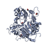
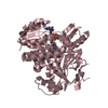
THE CONSTRUCT WAS EXPRESSED WITH A PURIFICATION TAG MGSDKIHHHHHHENLYFQG. THE TAG WAS REMOVED WITH ...THE CONSTRUCT WAS EXPRESSED WITH A PURIFICATION TAG MGSDKIHHHHHHENLYFQG. THE TAG WAS REMOVED WITH TEV PROTEASE LEAVING ONLY A GLYCINE (0) FOLLOWED BY THE TARGET SEQUENCE. THE CLONED CONSTRUCT CONTAINS RESIDUES 24-503 OF THE FULL LENGTH PROTEIN.
-
Experimental details
-
Experiment
Experiment
Method: X-RAY DIFFRACTION / Number of used crystals: 1
-
Sample preparation
Crystal
Density Matthews: 2.12 Å3/Da / Density % sol: 41.99 %
Crystal grow
Temperature: 277 K / Method: vapor diffusion, sitting drop / pH: 6.8 Details: 0.2000M NaNO3, 20.0000% PEG-3350, No Buffer pH 6.8, NANODROP, VAPOR DIFFUSION, SITTING DROP, temperature 277K
Monochromator: Single crystal Si(111) bent monochromator (horizontal focusing) Protocol: MAD / Monochromatic (M) / Laue (L): M / Scattering type: x-ray
Radiation wavelength
ID
Wavelength (Å)
Relative weight
1
0.91837
1
2
0.97848
1
3
0.97787
1
Reflection
Resolution: 1.35→29.656 Å / Num. obs: 87986 / % possible obs: 86.8 % / Redundancy: 4 % / Biso Wilson estimate: 12.865 Å2 / Rmerge(I) obs: 0.071 / Rsym value: 0.071 / Net I/σ(I): 12.5
Reflection shell
Resolution (Å)
Redundancy (%)
Rmerge(I) obs
Mean I/σ(I) obs
Num. measured all
Num. unique all
Rsym value
% possible all
1.35-1.39
2
0.259
2.6
13377
6668
0.259
91.1
1.39-1.42
2.2
0.232
3.1
14392
6610
0.232
90.8
1.42-1.46
4.1
0.304
2.3
26147
6424
0.304
90.4
1.46-1.51
4.1
0.24
2.9
25789
6265
0.24
90.3
1.51-1.56
4.1
0.201
3.5
24564
5944
0.201
89.4
1.56-1.61
4.2
0.169
4.2
24231
5782
0.169
89.1
1.61-1.67
4.2
0.143
4.9
23325
5547
0.143
88.5
1.67-1.74
4.3
0.125
5.6
22535
5297
0.125
88
1.74-1.82
4.3
0.11
6.1
21610
5042
0.11
87.1
1.82-1.91
4.3
0.097
6.8
20551
4747
0.097
86.7
1.91-2.01
4.4
0.087
7.5
19759
4528
0.087
85.8
2.01-2.13
4.4
0.079
7.9
18643
4220
0.079
85
2.13-2.28
4.5
0.071
8.8
17565
3928
0.071
84.2
2.28-2.46
4.5
0.063
10
16405
3624
0.063
83.4
2.46-2.7
4.6
0.062
10
15094
3293
0.062
82.2
2.7-3.02
4.7
0.058
10.7
13744
2947
0.058
81.2
3.02-3.49
4.7
0.058
10.8
12019
2558
0.058
79.9
3.49-4.27
4.7
0.049
12.8
9905
2130
0.049
78.5
4.27-6.04
4.6
0.049
12.9
7404
1607
0.049
76.4
6.04-29.67
4.3
0.056
11.8
3543
825
0.056
69.7
-
Phasing
Phasing
Method: MAD
-
Processing
Software
Name
Version
Classification
NB
REFMAC
5.2.0019
refinement
PHENIX
refinement
SHELX
phasing
MolProbity
3beta29
modelbuilding
SCALA
3.2.5
datascaling
PDB_EXTRACT
3.006
dataextraction
MOSFLM
datareduction
SHELXD
phasing
autoSHARP
phasing
Refinement
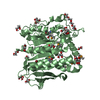
Method to determine structure: MAD / Resolution: 1.35→29.656 Å / Cor.coef. Fo:Fc: 0.976 / Cor.coef. Fo:Fc free: 0.97 / Occupancy max: 1 / Occupancy min: 0.25 / SU B: 0.773 / SU ML: 0.032 / Cross valid method: THROUGHOUT / σ(F): 0 / ESU R: 0.055 / ESU R Free: 0.056 Stereochemistry target values: MAXIMUM LIKELIHOOD WITH PHASES Details: HYDROGENS HAVE BEEN ADDED IN THE RIDING POSITIONS OTHER REFINEMENT REMARKS: 1.HYDROGENS HAVE BEEN ADDED IN THE RIDING POSITIONS. 2.A MET-INHIBITION PROTOCOL WAS USED FOR SELENOMETHIONINE ...Details: HYDROGENS HAVE BEEN ADDED IN THE RIDING POSITIONS OTHER REFINEMENT REMARKS: 1.HYDROGENS HAVE BEEN ADDED IN THE RIDING POSITIONS. 2.A MET-INHIBITION PROTOCOL WAS USED FOR SELENOMETHIONINE INCORPORATION DURING PROTEIN EXPRESSION. THE OCCUPANCY OF THE SE ATOMS IN THE MSE RESIDUES WAS REDUCED TO 0.75 TO ACCOUNT FOR THE REDUCED SCATTERING POWER DUE TO PARTIAL S-MET INCORPORATION. 3.CHLORIDE ANION FROM CRYSTALLIZATION AND ETHYLENE GLYCOL FROM CRYOPROTECTANT ARE MODELED IN THE STRUCTURE, RESPECTIVELY. 4.RESIDUE 191 IS CHEMICALLY MODIFIED TO S-HYDROXYCYSTINE WITH ELECTRON DENSITY SUPPORT.
Rfactor
Num. reflection
% reflection
Selection details
Rfree
0.167
4361
5 %
RANDOM
Rwork
0.146
-
-
-
obs
0.147
87959
86.51 %
-
Solvent computation
Ion probe radii: 0.8 Å / Shrinkage radii: 0.8 Å / VDW probe radii: 1.2 Å / Solvent model: BABINET MODEL WITH MASK
In the structure databanks used in Yorodumi, some data are registered as the other names, "COVID-19 virus" and "2019-nCoV". Here are the details of the virus and the list of structure data.
Jan 31, 2019. EMDB accession codes are about to change! (news from PDBe EMDB page)
EMDB accession codes are about to change! (news from PDBe EMDB page)
The allocation of 4 digits for EMDB accession codes will soon come to an end. Whilst these codes will remain in use, new EMDB accession codes will include an additional digit and will expand incrementally as the available range of codes is exhausted. The current 4-digit format prefixed with “EMD-” (i.e. EMD-XXXX) will advance to a 5-digit format (i.e. EMD-XXXXX), and so on. It is currently estimated that the 4-digit codes will be depleted around Spring 2019, at which point the 5-digit format will come into force.
The EM Navigator/Yorodumi systems omit the EMD- prefix.
Related info.:Q: What is EMD? / ID/Accession-code notation in Yorodumi/EM Navigator
Yorodumi is a browser for structure data from EMDB, PDB, SASBDB, etc.
This page is also the successor to EM Navigator detail page, and also detail information page/front-end page for Omokage search.
The word "yorodu" (or yorozu) is an old Japanese word meaning "ten thousand". "mi" (miru) is to see.
Related info.:EMDB / PDB / SASBDB / Comparison of 3 databanks / Yorodumi Search / Aug 31, 2016. New EM Navigator & Yorodumi / Yorodumi Papers / Jmol/JSmol / Function and homology information / Changes in new EM Navigator and Yorodumi
 Movie
Movie Controller

 Yorodumi
 Open data
 Basic information
 Components
 Keywords
 Function and homology information
 Bacteroides thetaiotaomicron VPI-5482 (bacteria)
 X-RAY DIFFRACTION /
  Authors
 Citation
 Structure visualization
 Downloads & links
 Other downloads


 PDBj
PDBj


 Assembly

 Mass: 35.453 Da / Num. of mol.: 1 / Source method: obtained synthetically / Formula: Cl
 Mass: 62.068 Da / Num. of mol.: 5 / Source method: obtained synthetically / Formula: C2H6O2
 Mass: 18.015 Da / Num. of mol.: 605 / Source method: isolated from a natural source / Formula: H2O
 Sample preparation
 / Beamline: BL11-1 / Wavelength: 0.91837,0.97848,0.97787
 Processing