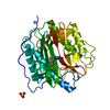
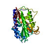
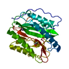
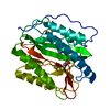
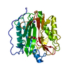
: / methionyl aminopeptidase / initiator methionyl aminopeptidase activity / cobalt ion binding / metalloaminopeptidase activity / protein processing / iron ion binding / metal ion binding / cytosol Similarity search - Function
Mass: 18.015 Da / Num. of mol.: 249 / Source method: isolated from a natural source / Formula: H2O
-
Experimental details
-
Experiment
Experiment
Method: X-RAY DIFFRACTION / Number of used crystals: 1
-
Sample preparation
Crystal
Density Matthews: 2.68 Å3/Da / Density % sol: 54.04 %
Crystal grow
Temperature: 298 K / Method: vapor diffusion, hanging drop / pH: 5.5 Details: 0.1 M Bis-Tris, pH 5.5, 1.1 M AMS, 50 mM NaCl, vapor diffusion, hanging drop, temperature 298K
Protocol: SINGLE WAVELENGTH / Monochromatic (M) / Laue (L): M / Scattering type: x-ray
Radiation wavelength
Wavelength: 1 Å / Relative weight: 1
Reflection
Redundancy: 10 % / Av σ(I) over netI: 43.78 / Number: 642578 / Rmerge(I) obs: 0.045 / Χ2: 0.66 / D res high: 1.4 Å / D res low: 50 Å / Num. obs: 64180 / % possible obs: 99.9
Diffraction reflection shell
Highest resolution (Å)
Lowest resolution (Å)
% possible obs (%)
ID
Rmerge(I) obs
Chi squared
Redundancy
3.8
50
98.8
1
0.024
0.476
10.6
3.02
3.8
99.7
1
0.03
0.732
7.6
2.63
3.02
100
1
0.036
0.735
9.6
2.39
2.63
100
1
0.037
0.675
10.2
2.22
2.39
100
1
0.06
1.203
10
2.09
2.22
100
1
0.041
0.578
10.3
1.99
2.09
100
1
0.045
0.531
10.4
1.9
1.99
100
1
0.07
0.983
10.3
1.83
1.9
100
1
0.067
0.732
10.3
1.76
1.83
100
1
0.069
0.639
10.3
1.71
1.76
100
1
0.075
0.553
10.3
1.66
1.71
100
1
0.081
0.529
10.3
1.62
1.66
100
1
0.092
0.547
10.3
1.58
1.62
100
1
0.103
0.555
10.3
1.54
1.58
100
1
0.114
0.553
10.3
1.51
1.54
100
1
0.126
0.585
10.3
1.48
1.51
100
1
0.149
0.627
10.3
1.45
1.48
100
1
0.16
0.573
10.2
1.42
1.45
100
1
0.184
0.658
9.8
1.4
1.42
100
1
0.215
0.745
8.3
Reflection
Resolution: 1.4→50 Å / Num. obs: 64180 / % possible obs: 99.9 % / Redundancy: 10 % / Rmerge(I) obs: 0.045 / Χ2: 0.657 / Net I/σ(I): 9.4
In the structure databanks used in Yorodumi, some data are registered as the other names, "COVID-19 virus" and "2019-nCoV". Here are the details of the virus and the list of structure data.
Jan 31, 2019. EMDB accession codes are about to change! (news from PDBe EMDB page)
EMDB accession codes are about to change! (news from PDBe EMDB page)
The allocation of 4 digits for EMDB accession codes will soon come to an end. Whilst these codes will remain in use, new EMDB accession codes will include an additional digit and will expand incrementally as the available range of codes is exhausted. The current 4-digit format prefixed with “EMD-” (i.e. EMD-XXXX) will advance to a 5-digit format (i.e. EMD-XXXXX), and so on. It is currently estimated that the 4-digit codes will be depleted around Spring 2019, at which point the 5-digit format will come into force.
The EM Navigator/Yorodumi systems omit the EMD- prefix.
Related info.:Q: What is EMD? / ID/Accession-code notation in Yorodumi/EM Navigator
Yorodumi is a browser for structure data from EMDB, PDB, SASBDB, etc.
This page is also the successor to EM Navigator detail page, and also detail information page/front-end page for Omokage search.
The word "yorodu" (or yorozu) is an old Japanese word meaning "ten thousand". "mi" (miru) is to see.
Related info.:EMDB / PDB / SASBDB / Comparison of 3 databanks / Yorodumi Search / Aug 31, 2016. New EM Navigator & Yorodumi / Yorodumi Papers / Jmol/JSmol / Function and homology information / Changes in new EM Navigator and Yorodumi
 Movie
Movie Controller


 Open data
 Basic information
 Components
 Keywords
 Function and homology information
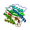
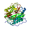
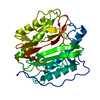
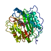
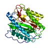
 Mycobacterium tuberculosis (bacteria)
 X-RAY DIFFRACTION /
  Authors
 Citation
 Structure visualization
 Downloads & links
 Other downloads
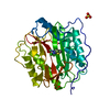

 PDBj
PDBj Assembly


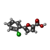


 Mass: 54.938 Da / Num. of mol.: 2 / Source method: obtained synthetically / Formula: Mn
 Mass: 222.624 Da / Num. of mol.: 2 / Source method: obtained synthetically / Formula: C11H7ClO3
 Mass: 96.063 Da / Num. of mol.: 1 / Source method: obtained synthetically / Formula: SO4
 Mass: 35.453 Da / Num. of mol.: 1 / Source method: obtained synthetically / Formula: Cl
 Sample preparation
 / Beamline: 19-BM / Wavelength: 1 Å
 Processing