 Movie
Movie Controller

[English] 日本語
 Yorodumi
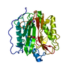
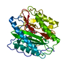
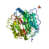
- PDB-1yj3: Crystal structure analysis of product bound methionine aminopepti... -
+ Open data

- Basic information
| Entry | Database: PDB / ID: 1yj3 | ||||||
|---|---|---|---|---|---|---|---|
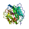
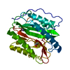
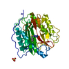
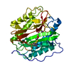
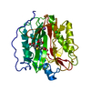
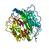
| Title | Crystal structure analysis of product bound methionine aminopeptidase Type 1c from Mycobacterium Tuberculosis | ||||||
 Components Components | Methionine aminopeptidase | ||||||
 Keywords Keywords | HYDROLASE / pita bread fold / complexed with Two Cobalt and Methionine / C285 modification by oxidized beta-mercapto ethanol | ||||||
| Function / homology |  Function and homology information Function and homology information: / methionyl aminopeptidase / initiator methionyl aminopeptidase activity / cobalt ion binding / metalloaminopeptidase activity / protein processing / iron ion binding / metal ion binding / cytosol Similarity search - Function | ||||||
| Biological species |   Mycobacterium tuberculosis (bacteria) Mycobacterium tuberculosis (bacteria) | ||||||
| Method |  X-RAY DIFFRACTION / MOLECULAR REPLACEMENT / Resolution: 1.6 Å X-RAY DIFFRACTION / MOLECULAR REPLACEMENT / Resolution: 1.6 Å | ||||||
 Authors Authors | Addlagatta, A. / Quillin, M.L. / Omotoso, O. / Liu, J.O. / Matthews, B.W. | ||||||
 Citation Citation | Journal: Biochemistry / Year: 2005 Title: Identification of an SH3-binding motif in a new class of methionine aminopeptidases from Mycobacterium tuberculosis suggests a mode of interaction with the ribosome. Authors: Addlagatta, A. / Quillin, M.L. / Omotoso, O. / Liu, J.O. / Matthews, B.W. | ||||||
| History |
|
- Structure visualization
| Structure viewer | Molecule: MolmilJmol/JSmol |
|---|
- Downloads & links
-Download
| PDBx/mmCIF format | 1yj3.cif.gz | 76.9 KB | Display | PDBx/mmCIF format |
|---|---|---|---|---|
| PDB format | pdb1yj3.ent.gz | 55.3 KB | Display | PDB format |
| PDBx/mmJSON format | 1yj3.json.gz | Tree view | PDBx/mmJSON format | |
| Others |  Other downloads Other downloads |
-Validation report
| Arichive directory | https://data.pdbj.org/pub/pdb/validation_reports/yj/1yj3ftp://data.pdbj.org/pub/pdb/validation_reports/yj/1yj3 | HTTPS FTP |
|---|
-Related structure data
| Related structure data | 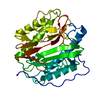 1y1nC  1c21S C: citing same article ( S: Starting model for refinement |
|---|---|
| Similar structure data |

-Links
 PDBj
PDBj


- Assembly
| Deposited unit | 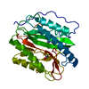
| ||||||||
|---|---|---|---|---|---|---|---|---|---|
| 1 |
| ||||||||
| Unit cell |
|
-Components
-Protein , 1 types, 1 molecules A
| #1: Protein | Mass: 31750.783 Da / Num. of mol.: 1 Source method: isolated from a genetically manipulated source Source: (gene. exp.) Mycobacterium tuberculosis (bacteria) / Gene: map, mapB / Plasmid: pET28a / Species (production host): Escherichia coli / Production host: References: UniProt: P0A5J2, UniProt: P9WK19*PLUS, methionyl aminopeptidase |
|---|
-Non-polymers , 5 types, 326 molecules 


| #2: Chemical |  Mass: 58.933 Da / Num. of mol.: 2 / Source method: obtained synthetically / Formula: Co Mass: 58.933 Da / Num. of mol.: 2 / Source method: obtained synthetically / Formula: Co#3: Chemical | ChemComp-K / |  Mass: 39.098 Da / Num. of mol.: 1 / Source method: obtained synthetically / Formula: K Mass: 39.098 Da / Num. of mol.: 1 / Source method: obtained synthetically / Formula: K#4: Chemical | ChemComp-MET / |  Type: L-peptide linking / Mass: 149.211 Da / Num. of mol.: 1 / Source method: obtained synthetically / Formula: C5H11NO2S Type: L-peptide linking / Mass: 149.211 Da / Num. of mol.: 1 / Source method: obtained synthetically / Formula: C5H11NO2S#5: Chemical | ChemComp-BME / |  Mass: 78.133 Da / Num. of mol.: 1 / Source method: obtained synthetically / Formula: C2H6OS Mass: 78.133 Da / Num. of mol.: 1 / Source method: obtained synthetically / Formula: C2H6OS#6: Water | ChemComp-HOH / | Mass: 18.015 Da / Num. of mol.: 321 / Source method: isolated from a natural source / Formula: H2O |
|---|
-Experimental details
-Experiment
| Experiment | Method: X-RAY DIFFRACTION / Number of used crystals: 1 |
|---|
- Sample preparation
| Crystal | Density Matthews: 2.11 Å3/Da / Density % sol: 41.66 % |
|---|---|
| Crystal grow | Temperature: 298 K / Method: vapor diffusion, hanging drop / pH: 6.5 Details: PEG 2000 monomethyl ether, BisTris, Oxidized beta-mercaptoethanol, pH 6.5, VAPOR DIFFUSION, HANGING DROP, temperature 298.0K |
-Data collection
| Diffraction | Mean temperature: 100 K |
|---|---|
| Diffraction source | Source: ROTATING ANODE / Type: RIGAKU / Wavelength: 1.5418 / Wavelength: 1.5418 Å |
| Detector | Type: RIGAKU RAXIS IV / Detector: IMAGE PLATE / Date: Nov 20, 2003 |
| Radiation | Monochromator: Ni MIRROR + Ni FILTER / Protocol: SINGLE WAVELENGTH / Monochromatic (M) / Laue (L): M / Scattering type: x-ray |
| Radiation wavelength | Wavelength: 1.5418 Å / Relative weight: 1 |
| Reflection | Resolution: 1.6→50 Å / Num. all: 37072 / Num. obs: 33032 / % possible obs: 94.2 % / Observed criterion σ(F): 0 / Observed criterion σ(I): 0 / Rsym value: 0.087 |
| Reflection shell | Resolution: 1.6→1.66 Å / Redundancy: 3.5 % / Rmerge(I) obs: 0.352 / Mean I/σ(I) obs: 1.8 / Num. unique all: 2979 / % possible all: 85.8 |
- Processing
| Software |
| ||||||||||||||||||||
|---|---|---|---|---|---|---|---|---|---|---|---|---|---|---|---|---|---|---|---|---|---|
| Refinement | Method to determine structure: MOLECULAR REPLACEMENT Starting model: PDB ENTRY 1C21 Resolution: 1.6→50 Å / Cross valid method: THROUGHOUT / σ(F): 0 / Stereochemistry target values: Engh & Huber Details: This sequence occurs naturally in Mycobacterium Tuberculosis.
| ||||||||||||||||||||
| Refinement step | Cycle: LAST / Resolution: 1.6→50 Å
|
