 Movie
Movie Controller

[English] 日本語
 Yorodumi
- PDB-3ipm: Crystal Structure of Archaeal 20S Proteasome in Complex with the ... -
+ Open data

- Basic information
| Entry | Database: PDB / ID: 3ipm | ||||||
|---|---|---|---|---|---|---|---|
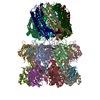
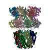
| Title | Crystal Structure of Archaeal 20S Proteasome in Complex with the C-terminus of PAN | ||||||
 Components Components |
| ||||||
 Keywords Keywords | HYDROLASE/HYDROLASE ACTIVATOR / Proteasome / proteasomal ATPase / protein degradation / AAA ATPase / electron cryomicroscopy / Hydrolase / Protease / Threonine protease / HYDROLASE-HYDROLASE ACTIVATOR COMPLEX | ||||||
| Function / homology |  Function and homology information Function and homology informationproteasome-activating nucleotidase complex / CTPase activity / proteasome activator complex / proteasome-activating activity / proteasome regulatory particle, base subcomplex / protein unfolding / proteasome endopeptidase complex / proteasome core complex, beta-subunit complex / threonine-type endopeptidase activity / proteasome core complex, alpha-subunit complex ...proteasome-activating nucleotidase complex / CTPase activity / proteasome activator complex / proteasome-activating activity / proteasome regulatory particle, base subcomplex / protein unfolding / proteasome endopeptidase complex / proteasome core complex, beta-subunit complex / threonine-type endopeptidase activity / proteasome core complex, alpha-subunit complex / proteasomal protein catabolic process / regulation of proteasomal protein catabolic process / ubiquitin-dependent protein catabolic process / endopeptidase activity / proteasome-mediated ubiquitin-dependent protein catabolic process / GTPase activity / ATP hydrolysis activity / proteolysis / ATP binding / cytoplasm Similarity search - Function | ||||||
| Biological species |   Thermoplasma acidophilum (acidophilic) Thermoplasma acidophilum (acidophilic) Methanocaldococcus jannaschii (archaea) Methanocaldococcus jannaschii (archaea) | ||||||
| Method |  X-RAY DIFFRACTION / SYNCHROTRON / MOLECULAR REPLACEMENT / Resolution: 4 Å X-RAY DIFFRACTION / SYNCHROTRON / MOLECULAR REPLACEMENT / Resolution: 4 Å | ||||||
 Authors Authors | Yu, Y. / Cheng, Y. | ||||||
 Citation Citation | Journal: EMBO J / Year: 2010 Title: Interactions of PAN's C-termini with archaeal 20S proteasome and implications for the eukaryotic proteasome-ATPase interactions. Authors: Yadong Yu / David M Smith / Ho Min Kim / Victor Rodriguez / Alfred L Goldberg / Yifan Cheng /  Abstract: Protein degradation in the 20S proteasome is regulated in eukaryotes by the 19S ATPase complex and in archaea by the homologous PAN ATPase ring complex. Subunits of these hexameric ATPases contain on ...Protein degradation in the 20S proteasome is regulated in eukaryotes by the 19S ATPase complex and in archaea by the homologous PAN ATPase ring complex. Subunits of these hexameric ATPases contain on their C-termini a conserved hydrophobic-tyrosine-X (HbYX) motif that docks into pockets in the 20S to stimulate the opening of a gated substrate entry channel. Here, we report the crystal structure of the archaeal 20S proteasome in complex with the C-terminus of the archaeal proteasome regulatory ATPase, PAN. This structure defines the detailed interactions between the critical C-terminal HbYX motif and the 20S alpha-subunits and indicates that the intersubunit pocket in the 20S undergoes an induced-fit conformational change on binding of the HbYX motif. This structure together with related mutagenesis data suggest how in eukaryotes certain proteasomal ATPases bind to specific pockets in an asymmetrical manner to regulate gate opening. | ||||||
| History |
|
- Structure visualization
| Structure viewer | Molecule: MolmilJmol/JSmol |
|---|
- Downloads & links
-Download
| PDBx/mmCIF format | 3ipm.cif.gz | 843 KB | Display | PDBx/mmCIF format |
|---|---|---|---|---|
| PDB format | pdb3ipm.ent.gz | 702.4 KB | Display | PDB format |
| PDBx/mmJSON format | 3ipm.json.gz | Tree view | PDBx/mmJSON format | |
| Others |  Other downloads Other downloads |
-Validation report
| Summary document | 3ipm_validation.pdf.gz | 596.3 KB | Display | wwPDB validaton report |
|---|---|---|---|---|
| Full document | 3ipm_full_validation.pdf.gz | 855.1 KB | Display | |
| Data in XML | 3ipm_validation.xml.gz | 183.1 KB | Display | |
| Data in CIF | 3ipm_validation.cif.gz | 237.6 KB | Display | |
| Arichive directory | https://data.pdbj.org/pub/pdb/validation_reports/ip/3ipmftp://data.pdbj.org/pub/pdb/validation_reports/ip/3ipm | HTTPS FTP |
-Related structure data
| Related structure data | 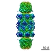 5130C 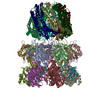 1ya7S C: citing same article ( S: Starting model for refinement |
|---|---|
| Similar structure data |


-Links
 PDBj
PDBj


- Assembly
| Deposited unit | 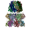
| ||||||||
|---|---|---|---|---|---|---|---|---|---|
| 1 | 
| ||||||||
| Unit cell |
|
-Components
| #1: Protein | Mass: 25829.447 Da / Num. of mol.: 7 Source method: isolated from a genetically manipulated source Source: (gene. exp.) Thermoplasma acidophilum (acidophilic) / Gene: psmA, Ta1288 / Plasmid: pRSET-A / Production host:  References: UniProt: P25156, proteasome endopeptidase complex #2: Protein | Mass: 23998.691 Da / Num. of mol.: 7 Source method: isolated from a genetically manipulated source Source: (gene. exp.) Thermoplasma acidophilum (acidophilic) / Gene: psmB, Ta0612 / Plasmid: pRSET-A / Production host: References: UniProt: P28061, proteasome endopeptidase complex #3: Protein | Mass: 26327.992 Da / Num. of mol.: 7 / Fragment: PA26 residues 2-223, PAN residues 424-430 / Mutation: E102A Source method: isolated from a genetically manipulated source Source: (gene. exp.) Methanocaldococcus jannaschii (archaea)Gene: Tb10.70.3660 / Plasmid: pET15b / Production host: |
|---|
-Experimental details
-Experiment
| Experiment | Method: X-RAY DIFFRACTION / Number of used crystals: 1 |
|---|
- Sample preparation
| Crystal | Density Matthews: 2.75 Å3/Da / Density % sol: 55.33 % |
|---|---|
| Crystal grow | Temperature: 298 K / Method: vapor diffusion, hanging drop / pH: 6.8 Details: 0.1 M MES pH6.8, 20% PEG 1500, VAPOR DIFFUSION, HANGING DROP, temperature 298K |
-Data collection
| Diffraction | Mean temperature: 100 K |
|---|---|
| Diffraction source | Source: SYNCHROTRON / Site: ALS / Beamline: 8.3.1 / Wavelength: 1.1159 Å |
| Detector | Type: ADSC QUANTUM 315 / Detector: CCD / Date: Dec 6, 2007 |
| Radiation | Monochromator: Double flat crystal, Si(111) / Protocol: SINGLE WAVELENGTH / Monochromatic (M) / Laue (L): M / Scattering type: x-ray |
| Radiation wavelength | Wavelength: 1.1159 Å / Relative weight: 1 |
| Reflection | Resolution: 4→89.5 Å / Num. all: 50164 / Num. obs: 48850 / % possible obs: 98.4 % / Observed criterion σ(F): 1 / Observed criterion σ(I): 1 / Redundancy: 3.2 % / Biso Wilson estimate: 53.1 Å2 / Rmerge(I) obs: 0.226 / Net I/σ(I): 2.9 |
| Reflection shell | Resolution: 4→4.22 Å / Redundancy: 3.3 % / Rmerge(I) obs: 0.449 / Mean I/σ(I) obs: 2.7 / Num. unique all: 7045 / % possible all: 98.6 |
- Processing
| Software |
| |||||||||||||||||||||||||
|---|---|---|---|---|---|---|---|---|---|---|---|---|---|---|---|---|---|---|---|---|---|---|---|---|---|---|
| Refinement | Method to determine structure: MOLECULAR REPLACEMENT Starting model: PDB entry 1YA7 Resolution: 4→89.51 Å / Isotropic thermal model: isotropic / Cross valid method: THROUGHOUT / σ(F): 0 / Stereochemistry target values: Engh & Huber
| |||||||||||||||||||||||||
| Displacement parameters | Biso mean: 92.7 Å2 | |||||||||||||||||||||||||
| Refine analyze |
| |||||||||||||||||||||||||
| Refinement step | Cycle: LAST / Resolution: 4→89.51 Å
| |||||||||||||||||||||||||
| Refine LS restraints |
| |||||||||||||||||||||||||
| LS refinement shell | Resolution: 4→4.14 Å / Rfactor Rfree error: 0.022
|
