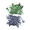
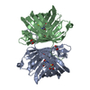
 ムービー
ムービー コントローラー


+ データを開く

- 基本情報
基本情報
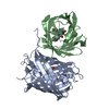
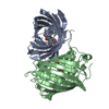
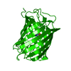
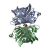
| 登録情報 | データベース: PDB / ID: 3ip2 | ||||||
|---|---|---|---|---|---|---|---|
| タイトル | Crystal structure of red fluorescent protein Neptune at pH 7.0 | ||||||
 要素 要素 | Neptune red fluorescent protein | ||||||
 キーワード キーワード | FLUORESCENT PROTEIN | ||||||
| 機能・相同性 | Green Fluorescent Protein / Green fluorescent protein / Green fluorescent protein-related / Green fluorescent protein / Green fluorescent protein / bioluminescence / Beta Barrel / Mainly Beta / Red fluorescent protein eqFP611 機能・相同性情報 機能・相同性情報 | ||||||
| 生物種 |  Entacmaea quadricolor (イソギンチャク) Entacmaea quadricolor (イソギンチャク) | ||||||
| 手法 |  X線回折 / シンクロトロン / 分子置換 / 解像度: 1.6 Å X線回折 / シンクロトロン / 分子置換 / 解像度: 1.6 Å | ||||||
 データ登録者 データ登録者 | Lin, M.Z. / McKeown, M.R. / Ng, H.L. / Aguilera, T.A. / Shaner, N.C. / Ma, W. / Adams, S.R. / Campbell, R.E. / Alber, T. / Tsien, R.Y. | ||||||
 引用 引用 | ジャーナル: Chem.Biol. / 年: 2009 タイトル: Autofluorescent proteins with excitation in the optical window for intravital imaging in mammals. 著者: Lin, M.Z. / McKeown, M.R. / Ng, H.L. / Aguilera, T.A. / Shaner, N.C. / Campbell, R.E. / Adams, S.R. / Gross, L.A. / Ma, W. / Alber, T. / Tsien, R.Y. | ||||||
| 履歴 |
|
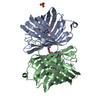
- 構造の表示
構造の表示
| 構造ビューア | 分子: MolmilJmol/JSmol |
|---|
- ダウンロードとリンク
-ダウンロード
| PDBx/mmCIF形式 | 3ip2.cif.gz | 116.6 KB | 表示 | PDBx/mmCIF形式 |
|---|---|---|---|---|
| PDB形式 | pdb3ip2.ent.gz | 90 KB | 表示 | PDB形式 |
| PDBx/mmJSON形式 | 3ip2.json.gz | ツリー表示 | PDBx/mmJSON形式 | |
| その他 |  その他のダウンロード その他のダウンロード |
-検証レポート
| 文書・要旨 | 3ip2_validation.pdf.gz | 428.7 KB | 表示 | wwPDB検証レポート |
|---|---|---|---|---|
| 文書・詳細版 | 3ip2_full_validation.pdf.gz | 431.5 KB | 表示 | |
| XML形式データ | 3ip2_validation.xml.gz | 13.7 KB | 表示 | |
| CIF形式データ | 3ip2_validation.cif.gz | 19.7 KB | 表示 | |
| アーカイブディレクトリ | https://data.pdbj.org/pub/pdb/validation_reports/ip/3ip2ftp://data.pdbj.org/pub/pdb/validation_reports/ip/3ip2 | HTTPS FTP |
-関連構造データ
| 関連構造データ |  3bxaS S: 精密化の開始モデル |
|---|---|
| 類似構造データ |


-リンク
 PDBj
PDBj

- 集合体
集合体
| 登録構造単位 | 
| ||||||||
|---|---|---|---|---|---|---|---|---|---|
| 1 | 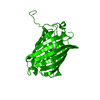
| ||||||||
| 単位格子 |
| ||||||||
| Components on special symmetry positions |
| ||||||||
| 詳細 | authors state that this is a WEAK DIMER (DISSOCIATION CONSTANT 500 NANOMOLAR). |
-要素
| #1: タンパク質 | 分子量: 27369.211 Da / 分子数: 1 / 由来タイプ: 組換発現 由来: (組換発現) Entacmaea quadricolor (イソギンチャク)発現宿主:  |
|---|---|
| #2: 水 | ChemComp-HOH /  分子量: 18.015 Da / 分子数: 212 / 由来タイプ: 天然 / 式: H2O 分子量: 18.015 Da / 分子数: 212 / 由来タイプ: 天然 / 式: H2O |
| 配列の詳細 | RESIDUES MET 63, TYR 64 AND GLY 65 AUTOCATALYTICALLY FORM THE CHROMOPHORE NRQ LABELLED AS RESIDUE ...RESIDUES MET 63, TYR 64 AND GLY 65 AUTOCATALY |
-実験情報
-実験
| 実験 | 手法: X線回折 / 使用した結晶の数: 1 |
|---|
- 試料調製
試料調製
| 結晶 | マシュー密度: 2.06 Å3/Da / 溶媒含有率: 40.39 % |
|---|---|
| 結晶化 | 温度: 291 K / 手法: 蒸気拡散法, シッティングドロップ法 / pH: 7 詳細: 1 uL of protein at 10 mg/mL was mixed with 1 uL of 0.1 M HEPES, 0.2 M CaCl2, 20% PEG 6000, pH 7.0, VAPOR DIFFUSION, SITTING DROP, temperature 291K |
-データ収集
| 回折 | 平均測定温度: 100 K |
|---|---|
| 放射光源 | 由来: シンクロトロン / サイト: ALS  / ビームライン: 8.3.1 / 波長: 1.11586 Å / ビームライン: 8.3.1 / 波長: 1.11586 Å |
| 検出器 | タイプ: ADSC QUANTUM 315r / 検出器: CCD / 日付: 2009年4月1日 |
| 放射 | モノクロメーター: KHOZU Double flat crystal / プロトコル: SINGLE WAVELENGTH / 単色(M)・ラウエ(L): M / 散乱光タイプ: x-ray |
| 放射波長 | 波長: 1.11586 Å / 相対比: 1 |
| 反射 | 解像度: 1.6→92.1 Å / Num. all: 30810 / Num. obs: 30755 / % possible obs: 99.9 % / Observed criterion σ(F): -3 / Observed criterion σ(I): -3 / 冗長度: 8.5 % / Rsym value: 0.055 / Net I/σ(I): 23.8 |
| 反射 シェル | 解像度: 1.6→1.68 Å / Mean I/σ(I) obs: 2.2 / Num. unique all: 4369 / Rsym value: 0.778 / % possible all: 99.7 |
- 解析
解析
| ソフトウェア |
| ||||||||||||||||||||||||||||||||||||||||||||||||||||||||||||||||||||||||||||||||||||
|---|---|---|---|---|---|---|---|---|---|---|---|---|---|---|---|---|---|---|---|---|---|---|---|---|---|---|---|---|---|---|---|---|---|---|---|---|---|---|---|---|---|---|---|---|---|---|---|---|---|---|---|---|---|---|---|---|---|---|---|---|---|---|---|---|---|---|---|---|---|---|---|---|---|---|---|---|---|---|---|---|---|---|---|---|---|
| 精密化 | 構造決定の手法: 分子置換 開始モデル: pdb entry 3BXA 解像度: 1.6→65.2 Å / SU ML: 0.27 / σ(F): -3 / 立体化学のターゲット値: Engh & Huber / 詳細: TLS refinement
| ||||||||||||||||||||||||||||||||||||||||||||||||||||||||||||||||||||||||||||||||||||
| 溶媒の処理 | 減衰半径: 0.9 Å / VDWプローブ半径: 1.11 Å / 溶媒モデル: FLAT BULK SOLVENT MODEL / Bsol: 60.022 Å2 / ksol: 0.42 e/Å3 | ||||||||||||||||||||||||||||||||||||||||||||||||||||||||||||||||||||||||||||||||||||
| 精密化ステップ | サイクル: LAST / 解像度: 1.6→65.2 Å
| ||||||||||||||||||||||||||||||||||||||||||||||||||||||||||||||||||||||||||||||||||||
| LS精密化 シェル |
| ||||||||||||||||||||||||||||||||||||||||||||||||||||||||||||||||||||||||||||||||||||
| 精密化 TLS | 手法: refined / Origin x: -0.8391 Å / Origin y: 21.7715 Å / Origin z: 25.2752 Å
| ||||||||||||||||||||||||||||||||||||||||||||||||||||||||||||||||||||||||||||||||||||
| 精密化 TLSグループ | Selection details: chain A |
