Single helix bin / Electron Transport, Fmn-binding Protein; Chain A / Pnp Oxidase; Chain A / FMN-binding split barrel / Single alpha-helices involved in coiled-coils or other helix-helix interfaces / Roll / Up-down Bundle / Mainly Beta / Mainly Alpha Similarity search - Domain/homology
Component-ID: 1 / Ens-ID: 1 / Beg auth comp-ID: VAL / Beg label comp-ID: VAL / End auth comp-ID: ARG / End label comp-ID: ARG / Refine code: 6 / Auth seq-ID: 5 - 146 / Label seq-ID: 6 - 147
Dom-ID
Auth asym-ID
Label asym-ID
1
A
A
2
B
B
Details
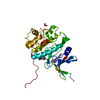
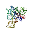
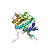
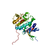
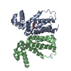
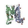
CRYSTAL PACKING ANALYSIS AND ANALYTICAL SIZE EXCLUSION CHROMATOGRAPHY SUPPORT THE ASSIGNMENT OF A DIMER AS THE SIGNIFICANT OLIGOMERIC FORM IN SOLUTION.
-
Components
#1: Protein
FMN-bindingprotein
Mass: 17184.330 Da / Num. of mol.: 2 Source method: isolated from a genetically manipulated source Source: (gene. exp.) Syntrophomonas wolfei subsp. wolfei (bacteria) Strain: Goettingen / Gene: Swol_0183, YP_752906.1 / Plasmid: SpeedET / Production host: Escherichia coli (E. coli) / Strain (production host): HK100 / References: UniProt: Q0B0G9
Mass: 18.015 Da / Num. of mol.: 41 / Source method: isolated from a natural source / Formula: H2O
Has protein modification
Y
Sequence details
THIS CONSTRUCT WAS EXPRESSED WITH A PURIFICATION TAG MGSDKIHHHHHHENLYFQG. THE TAG WAS REMOVED WITH ...THIS CONSTRUCT WAS EXPRESSED WITH A PURIFICATION TAG MGSDKIHHHHHHENLYFQG. THE TAG WAS REMOVED WITH TEV PROTEASE LEAVING ONLY A GLYCINE (0) FOLLOWED BY THE TARGET SEQUENCE.
-
Experimental details
-
Experiment
Experiment
Method: X-RAY DIFFRACTION / Number of used crystals: 1
-
Sample preparation
Crystal
Density Matthews: 2.31 Å3/Da / Density % sol: 46.79 %
Crystal grow
Temperature: 277 K / Method: vapor diffusion, sitting drop / pH: 4.29 Details: 30.5000% 2-methyl-2,4-pentanediol, 0.1M sodium acetate pH 4.29, NANODROP, VAPOR DIFFUSION, SITTING DROP, temperature 277K
Resolution: 2.12→28.712 Å / Num. obs: 18788 / % possible obs: 99.5 % / Observed criterion σ(I): -3 / Redundancy: 7.12 % / Biso Wilson estimate: 35.044 Å2 / Rmerge(I) obs: 0.057 / Net I/σ(I): 16.01
Reflection shell
Resolution (Å)
Rmerge(I) obs
Mean I/σ(I) obs
Num. measured obs
Num. unique obs
Diffraction-ID
% possible all
2.12-2.2
0.554
2.5
13800
3674
1
99.5
2.2-2.28
0.461
3
12044
3141
1
99.1
2.28-2.39
0.385
3.6
14213
3692
1
98.9
2.39-2.51
0.289
4.7
12709
3303
1
99.4
2.51-2.67
0.2
6.7
13592
3530
1
99.6
2.67-2.88
0.142
9.4
13648
3537
1
99.5
2.88-3.16
0.085
14.7
13141
3396
1
99.8
3.16-3.62
0.046
25.4
13641
3533
1
99.7
3.62-4.55
0.027
39.5
13429
3477
1
99.8
4.55-28.712
0.02
49.8
13623
3538
1
99.3
-
Phasing
Phasing
Method: MAD
-
Processing
Software
Name
Version
Classification
NB
REFMAC
5.5.0053
refinement
PHENIX
refinement
SHELX
phasing
MolProbity
3beta29
modelbuilding
XSCALE
datascaling
PDB_EXTRACT
3.006
dataextraction
XDS
datareduction
SHARP
phasing
Refinement
Method to determine structure: MAD / Resolution: 2.12→28.712 Å / Cor.coef. Fo:Fc: 0.948 / Cor.coef. Fo:Fc free: 0.936 / Occupancy max: 1 / Occupancy min: 0.4 / SU B: 13.723 / SU ML: 0.166 / TLS residual ADP flag: LIKELY RESIDUAL / Cross valid method: THROUGHOUT / σ(F): 0 / ESU R: 0.257 / ESU R Free: 0.202 Stereochemistry target values: MAXIMUM LIKELIHOOD WITH PHASES Details: 1.HYDROGENS HAVE BEEN ADDED IN THE RIDING POSITIONS. 2.ATOM RECORDS CONTAIN RESIDUAL B FACTORS ONLY. 3.A MET-INHIBITION PROTOCOL WAS USED FOR SELENOMETHIONINE INCORPORATION DURING PROTEIN ...Details: 1.HYDROGENS HAVE BEEN ADDED IN THE RIDING POSITIONS. 2.ATOM RECORDS CONTAIN RESIDUAL B FACTORS ONLY. 3.A MET-INHIBITION PROTOCOL WAS USED FOR SELENOMETHIONINE INCORPORATION DURING PROTEIN EXPRESSION. THE OCCUPANCY OF THE SE ATOMS IN THE MSE RESIDUES WAS REDUCED TO 0.75 FOR THE REDUCED SCATTERING POWER DUE TO PARTIAL S-MET INCORPORATION. 4.ENDOGENOUS FLAVIN MONONUCLEOTIDE(FMN) HAS BEEN MODELED AT THE PUTATIVE ACTIVE SITE BASED ON CLEAR ELECTRON DENSITY. 5.2,4-METHANEPENTANEDIOL (MPD) FROM THE CRYSTALLIZATION SOLUTION HAS BEEN MODELED IN THE SOLVENT STRUCTURE.
Rfactor
Num. reflection
% reflection
Selection details
Rfree
0.254
958
5.1 %
RANDOM
Rwork
0.219
-
-
-
obs
0.221
18752
99.54 %
-
Solvent computation
Ion probe radii: 0.8 Å / Shrinkage radii: 0.8 Å / VDW probe radii: 1.2 Å / Solvent model: MASK
In the structure databanks used in Yorodumi, some data are registered as the other names, "COVID-19 virus" and "2019-nCoV". Here are the details of the virus and the list of structure data.
Jan 31, 2019. EMDB accession codes are about to change! (news from PDBe EMDB page)
EMDB accession codes are about to change! (news from PDBe EMDB page)
The allocation of 4 digits for EMDB accession codes will soon come to an end. Whilst these codes will remain in use, new EMDB accession codes will include an additional digit and will expand incrementally as the available range of codes is exhausted. The current 4-digit format prefixed with “EMD-” (i.e. EMD-XXXX) will advance to a 5-digit format (i.e. EMD-XXXXX), and so on. It is currently estimated that the 4-digit codes will be depleted around Spring 2019, at which point the 5-digit format will come into force.
The EM Navigator/Yorodumi systems omit the EMD- prefix.
Related info.:Q: What is EMD? / ID/Accession-code notation in Yorodumi/EM Navigator
Yorodumi is a browser for structure data from EMDB, PDB, SASBDB, etc.
This page is also the successor to EM Navigator detail page, and also detail information page/front-end page for Omokage search.
The word "yorodu" (or yorozu) is an old Japanese word meaning "ten thousand". "mi" (miru) is to see.
Related info.:EMDB / PDB / SASBDB / Comparison of 3 databanks / Yorodumi Search / Aug 31, 2016. New EM Navigator & Yorodumi / Yorodumi Papers / Jmol/JSmol / Function and homology information / Changes in new EM Navigator and Yorodumi
 Movie
Movie Controller

 Yorodumi
 Open data
 Basic information
 Components
 Keywords
 Function and homology information
 Syntrophomonas wolfei subsp. wolfei (bacteria)
 X-RAY DIFFRACTION /
  Authors
 Citation
 Structure visualization
 Downloads & links
 Other downloads
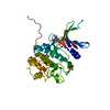

 PDBj
PDBj Assembly
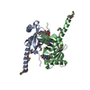

 Mass: 456.344 Da / Num. of mol.: 2 / Source method: obtained synthetically / Formula: C17H21N4O9P
 Mass: 118.174 Da / Num. of mol.: 1 / Source method: obtained synthetically / Formula: C6H14O2 / Comment: precipitant*YM
 Mass: 18.015 Da / Num. of mol.: 41 / Source method: isolated from a natural source / Formula: H2O
 Sample preparation
 / Beamline: BL9-2 / Wavelength: 0.91162,0.97936,0.97922
 Processing