- PDB-3in5: Structure of human DNA polymerase kappa inserting dATP opposite a... -
+
Open data
ID or keywords:
Loading...
-
Basic information
Entry
Database: PDB / ID: 3in5
Title
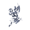
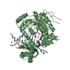
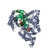
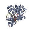
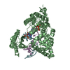
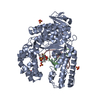
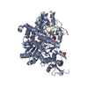
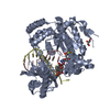
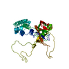
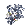
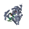
Structure of human DNA polymerase kappa inserting dATP opposite an 8-oxoG DNA lesion
Components
DNA (5'-D(*C*CP*TP*AP*(8OG)P*GP*AP*GP*TP*CP*CP*TP*TP*CP*CP*CP*CP*C)-3')
DNA (5'-D(*GP*G*GP*GP*GP*AP*AP*GP*GP*AP*CP*TP*(DOC))-3')
DNA polymerase kappa
Keywords
Transferase/DNA / Alternative splicing / DNA damage / DNA repair / DNA replication / DNA synthesis / DNA-binding / DNA-directed DNA polymerase / Magnesium / Metal-binding / Mutator protein / Nucleotidyltransferase / Nucleus / Phosphoprotein / Polymorphism / Schiff base / Transferase / Zinc / Zinc-finger / Transferase-DNA COMPLEX
Function / homology
Function and homology information
nucleotide-excision repair, DNA gap filling / DNA biosynthetic process / error-prone translesion synthesis / site of DNA damage / translesion synthesis / Translesion synthesis by POLK / Gap-filling DNA repair synthesis and ligation in GG-NER / Termination of translesion DNA synthesis / HDR through Homologous Recombination (HRR) / Dual Incision in GG-NER ...nucleotide-excision repair, DNA gap filling / DNA biosynthetic process / error-prone translesion synthesis / site of DNA damage / translesion synthesis / Translesion synthesis by POLK / Gap-filling DNA repair synthesis and ligation in GG-NER / Termination of translesion DNA synthesis / HDR through Homologous Recombination (HRR) / Dual Incision in GG-NER / cellular response to UV / Dual incision in TC-NER / Gap-filling DNA repair synthesis and ligation in TC-NER / DNA-directed DNA polymerase / damaged DNA binding / DNA-directed DNA polymerase activity / DNA repair / DNA damage response / zinc ion binding / nucleoplasm / nucleus Similarity search - Function
DNA polymerase; domain 1 - #810 / Rad18-like CCHC zinc finger / DNA polymerase type-Y, HhH motif / IMS family HHH motif / DNA polymerase IV / : / DNA polymerase, Y-family, little finger domain / MutS, DNA mismatch repair protein, domain I - #60 / Rad18, zinc finger UBZ4-type / Zinc finger UBZ4-type profile. ...DNA polymerase; domain 1 - #810 / Rad18-like CCHC zinc finger / DNA polymerase type-Y, HhH motif / IMS family HHH motif / DNA polymerase IV / : / DNA polymerase, Y-family, little finger domain / MutS, DNA mismatch repair protein, domain I - #60 / Rad18, zinc finger UBZ4-type / Zinc finger UBZ4-type profile. / MutS, DNA mismatch repair protein, domain I / DNA polymerase, Y-family, little finger domain / impB/mucB/samB family C-terminal domain / UmuC domain / DNA polymerase, Y-family, little finger domain superfamily / impB/mucB/samB family / UmuC domain profile. / Reverse transcriptase/Diguanylate cyclase domain / Dna Ligase; domain 1 / DNA polymerase; domain 1 / Reverse transcriptase/Diguanylate cyclase domain / Alpha-Beta Plaits / DNA/RNA polymerase superfamily / 2-Layer Sandwich / Orthogonal Bundle / 3-Layer(aba) Sandwich / Mainly Alpha / Alpha Beta Similarity search - Domain/homology
A: DNA polymerase kappa B: DNA polymerase kappa P: DNA (5'-D(*GP*G*GP*GP*GP*AP*AP*GP*GP*AP*CP*TP*(DOC))-3') T: DNA (5'-D(*C*CP*TP*AP*(8OG)P*GP*AP*GP*TP*CP*CP*TP*TP*CP*CP*CP*CP*C)-3') Q: DNA (5'-D(*GP*G*GP*GP*GP*AP*AP*GP*GP*AP*CP*TP*(DOC))-3') U: DNA (5'-D(*C*CP*TP*AP*(8OG)P*GP*AP*GP*TP*CP*CP*TP*TP*CP*CP*CP*CP*C)-3') hetero molecules
A: DNA polymerase kappa P: DNA (5'-D(*GP*G*GP*GP*GP*AP*AP*GP*GP*AP*CP*TP*(DOC))-3') T: DNA (5'-D(*C*CP*TP*AP*(8OG)P*GP*AP*GP*TP*CP*CP*TP*TP*CP*CP*CP*CP*C)-3') hetero molecules
B: DNA polymerase kappa Q: DNA (5'-D(*GP*G*GP*GP*GP*AP*AP*GP*GP*AP*CP*TP*(DOC))-3') U: DNA (5'-D(*C*CP*TP*AP*(8OG)P*GP*AP*GP*TP*CP*CP*TP*TP*CP*CP*CP*CP*C)-3') hetero molecules
Resolution: 3.2→47.12 Å / Cor.coef. Fo:Fc: 0.907 / Cor.coef. Fo:Fc free: 0.858 / SU B: 18.79 / SU ML: 0.326 / Cross valid method: THROUGHOUT / ESU R Free: 0.449 / Stereochemistry target values: MAXIMUM LIKELIHOOD / Details: HYDROGENS HAVE BEEN ADDED IN THE RIDING POSITIONS
Rfactor
Num. reflection
% reflection
Selection details
Rfree
0.27403
1640
5 %
RANDOM
Rwork
0.22453
-
-
-
obs
0.22696
31139
99.93 %
-
all
-
34388
-
-
Solvent computation
Ion probe radii: 0.8 Å / Shrinkage radii: 0.8 Å / VDW probe radii: 1.4 Å / Solvent model: MASK
Displacement parameters
Biso mean: 46.402 Å2
Baniso -1
Baniso -2
Baniso -3
1-
3.18 Å2
0 Å2
0 Å2
2-
-
1.33 Å2
0 Å2
3-
-
-
-4.51 Å2
Refinement step
Cycle: LAST / Resolution: 3.2→47.12 Å
Protein
Nucleic acid
Ligand
Solvent
Total
Num. atoms
6757
1056
64
18
7895
Refine LS restraints
Refine-ID
Type
Dev ideal
Dev ideal target
Number
X-RAY DIFFRACTION
r_bond_refined_d
0.012
0.022
8111
X-RAY DIFFRACTION
r_bond_other_d
0.002
0.02
5150
X-RAY DIFFRACTION
r_angle_refined_deg
1.567
2.144
11192
X-RAY DIFFRACTION
r_angle_other_deg
0.964
3
12639
X-RAY DIFFRACTION
r_dihedral_angle_1_deg
5.589
5
873
X-RAY DIFFRACTION
r_dihedral_angle_2_deg
37.408
24.5
300
X-RAY DIFFRACTION
r_dihedral_angle_3_deg
19.639
15
1241
X-RAY DIFFRACTION
r_dihedral_angle_4_deg
21.503
15
46
X-RAY DIFFRACTION
r_chiral_restr
0.069
0.2
1278
X-RAY DIFFRACTION
r_gen_planes_refined
0.005
0.02
8203
X-RAY DIFFRACTION
r_gen_planes_other
0.001
0.02
1447
X-RAY DIFFRACTION
r_nbd_refined
X-RAY DIFFRACTION
r_nbd_other
X-RAY DIFFRACTION
r_nbtor_refined
X-RAY DIFFRACTION
r_nbtor_other
X-RAY DIFFRACTION
r_xyhbond_nbd_refined
X-RAY DIFFRACTION
r_xyhbond_nbd_other
X-RAY DIFFRACTION
r_metal_ion_refined
X-RAY DIFFRACTION
r_metal_ion_other
X-RAY DIFFRACTION
r_symmetry_vdw_refined
X-RAY DIFFRACTION
r_symmetry_vdw_other
X-RAY DIFFRACTION
r_symmetry_hbond_refined
X-RAY DIFFRACTION
r_symmetry_hbond_other
X-RAY DIFFRACTION
r_symmetry_metal_ion_refined
X-RAY DIFFRACTION
r_symmetry_metal_ion_other
X-RAY DIFFRACTION
r_mcbond_it
0.514
1.5
4369
X-RAY DIFFRACTION
r_mcbond_other
0.059
1.5
1755
X-RAY DIFFRACTION
r_mcangle_it
0.979
2
7006
X-RAY DIFFRACTION
r_scbond_it
1.196
3
3742
X-RAY DIFFRACTION
r_scangle_it
2.131
4.5
4185
X-RAY DIFFRACTION
r_rigid_bond_restr
X-RAY DIFFRACTION
r_sphericity_free
X-RAY DIFFRACTION
r_sphericity_bonded
LS refinement shell
Resolution: 3.2→3.283 Å / Total num. of bins used: 20
Rfactor
Num. reflection
% reflection
Rfree
0.289
112
-
Rwork
0.283
2264
-
obs
-
-
99.87 %
+
About Yorodumi
-
News
-
Feb 9, 2022. New format data for meta-information of EMDB entries
New format data for meta-information of EMDB entries
Version 3 of the EMDB header file is now the official format.
The previous official version 1.9 will be removed from the archive.
In the structure databanks used in Yorodumi, some data are registered as the other names, "COVID-19 virus" and "2019-nCoV". Here are the details of the virus and the list of structure data.
Jan 31, 2019. EMDB accession codes are about to change! (news from PDBe EMDB page)
EMDB accession codes are about to change! (news from PDBe EMDB page)
The allocation of 4 digits for EMDB accession codes will soon come to an end. Whilst these codes will remain in use, new EMDB accession codes will include an additional digit and will expand incrementally as the available range of codes is exhausted. The current 4-digit format prefixed with “EMD-” (i.e. EMD-XXXX) will advance to a 5-digit format (i.e. EMD-XXXXX), and so on. It is currently estimated that the 4-digit codes will be depleted around Spring 2019, at which point the 5-digit format will come into force.
The EM Navigator/Yorodumi systems omit the EMD- prefix.
Related info.:Q: What is EMD? / ID/Accession-code notation in Yorodumi/EM Navigator
Yorodumi is a browser for structure data from EMDB, PDB, SASBDB, etc.
This page is also the successor to EM Navigator detail page, and also detail information page/front-end page for Omokage search.
The word "yorodu" (or yorozu) is an old Japanese word meaning "ten thousand". "mi" (miru) is to see.
Related info.:EMDB / PDB / SASBDB / Comparison of 3 databanks / Yorodumi Search / Aug 31, 2016. New EM Navigator & Yorodumi / Yorodumi Papers / Jmol/JSmol / Function and homology information / Changes in new EM Navigator and Yorodumi
 Movie
Movie Controller

 Yorodumi
 Open data
 Basic information
 Components
 Keywords
 Function and homology information
 Homo sapiens (human)
 X-RAY DIFFRACTION /
  Authors
 Citation
 Structure visualization
 Downloads & links
 Other downloads
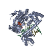

 PDBj
PDBj


 Assembly


 Mass: 507.181 Da / Num. of mol.: 2 / Source method: obtained synthetically / Formula: C10H16N5O13P3 / Comment: ATP, energy-carrying molecule*YM
 Mass: 24.305 Da / Num. of mol.: 4 / Source method: obtained synthetically / Formula: Mg
 Sample preparation
 / Beamline: X25 / Wavelength: 1.1 Å
 Processing