 Movie
Movie Controller

[English] 日本語
 Yorodumi
- PDB-3ikt: Crystal structure of a Rex-family repressor/DNA/NAD+ complex from... -
+ Open data

- Basic information
| Entry | Database: PDB / ID: 3ikt | ||||||
|---|---|---|---|---|---|---|---|
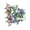
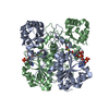
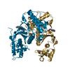
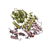
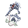
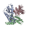
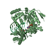
| Title | Crystal structure of a Rex-family repressor/DNA/NAD+ complex from Thermus aquaticus | ||||||
 Components Components |
| ||||||
 Keywords Keywords | DNA BINDING PROTEIN/DNA / redox-sensing / winged helix / Rossmann fold / Nicotinamide Adenine Dinucleotide / NAD / Rex / Thermus aquaticus / DNA BINDING PROTEIN / Cytoplasm / DNA-binding / Repressor / Transcription / Transcription regulation / DNA BINDING PROTEIN-DNA COMPLEX | ||||||
| Function / homology |  Function and homology information Function and homology informationresponse to redox state / DNA-binding transcription factor activity / negative regulation of DNA-templated transcription / DNA-templated transcription / DNA binding / cytoplasm Similarity search - Function | ||||||
| Biological species |   Thermus thermophilus HB27 (bacteria) Thermus thermophilus HB27 (bacteria) | ||||||
| Method |  X-RAY DIFFRACTION / SYNCHROTRON / MOLECULAR REPLACEMENT / Resolution: 2.26 Å X-RAY DIFFRACTION / SYNCHROTRON / MOLECULAR REPLACEMENT / Resolution: 2.26 Å | ||||||
 Authors Authors | McLaughlin, K.J. / Kielkopf, C.L. | ||||||
 Citation Citation | Journal: Mol.Cell / Year: 2010 Title: Structural basis for NADH/NAD+ redox sensing by a Rex family repressor. Authors: McLaughlin, K.J. / Strain-Damerell, C.M. / Xie, K. / Brekasis, D. / Soares, A.S. / Paget, M.S. / Kielkopf, C.L. | ||||||
| History |
|
- Structure visualization
| Structure viewer | Molecule: MolmilJmol/JSmol |
|---|
- Downloads & links
-Download
| PDBx/mmCIF format | 3ikt.cif.gz | 128.6 KB | Display | PDBx/mmCIF format |
|---|---|---|---|---|
| PDB format | pdb3ikt.ent.gz | 94.7 KB | Display | PDB format |
| PDBx/mmJSON format | 3ikt.json.gz | Tree view | PDBx/mmJSON format | |
| Others |  Other downloads Other downloads |
-Validation report
| Arichive directory | https://data.pdbj.org/pub/pdb/validation_reports/ik/3iktftp://data.pdbj.org/pub/pdb/validation_reports/ik/3ikt | HTTPS FTP |
|---|
-Related structure data
| Related structure data | 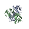 3ikvC  3il2C 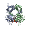 1xcbS S: Starting model for refinement C: citing same article ( |
|---|---|
| Similar structure data |

-Links
 PDBj
PDBj


- Assembly
| Deposited unit | 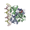
| ||||||||
|---|---|---|---|---|---|---|---|---|---|
| 1 |
| ||||||||
| Unit cell |
| ||||||||
| Details | BIOLOGICAL UNIT IS THE SAME AS ASYMMETRIC UNIT. |
-Components
| #1: Protein | Mass: 22730.438 Da / Num. of mol.: 2 Source method: isolated from a genetically manipulated source Source: (gene. exp.) Thermus thermophilus HB27 (bacteria) / Strain: HB27 / DSM 7039 / Gene: rex, TT_C1293 / Plasmid: pET24 / Production host: #2: DNA chain | Mass: 6753.354 Da / Num. of mol.: 2 / Source method: obtained synthetically / Details: Synthetic 22bp dsDNA obtained from IDT #3: Chemical | ChemComp-NAD / |   Mass: 663.425 Da / Num. of mol.: 1 / Source method: obtained synthetically / Formula: C21H27N7O14P2 / Comment: NAD*YM Mass: 663.425 Da / Num. of mol.: 1 / Source method: obtained synthetically / Formula: C21H27N7O14P2 / Comment: NAD*YM#4: Water | ChemComp-HOH / |  Mass: 18.015 Da / Num. of mol.: 378 / Source method: isolated from a natural source / Formula: H2O Mass: 18.015 Da / Num. of mol.: 378 / Source method: isolated from a natural source / Formula: H2O |
|---|
-Experimental details
-Experiment
| Experiment | Method: X-RAY DIFFRACTION / Number of used crystals: 1 |
|---|
- Sample preparation
| Crystal | Density Matthews: 2.93 Å3/Da / Density % sol: 57.98 % | ||||||||||||||||||||||||||||||||||||
|---|---|---|---|---|---|---|---|---|---|---|---|---|---|---|---|---|---|---|---|---|---|---|---|---|---|---|---|---|---|---|---|---|---|---|---|---|---|
| Crystal grow | Temperature: 298 K / Method: vapor diffusion, hanging drop / pH: 6 Details: 20-30% PEG 400, 0.1M Na Cacodylate pH 6.0, 0.2M CaCl2, 5-10% Glycerol, VAPOR DIFFUSION, HANGING DROP, temperature 298K | ||||||||||||||||||||||||||||||||||||
| Components of the solutions |
|
-Data collection
| Diffraction | Mean temperature: 100 K |
|---|---|
| Diffraction source | Source: SYNCHROTRON / Site: NSLS  / Beamline: X12B / Wavelength: 1 Å / Beamline: X12B / Wavelength: 1 Å |
| Detector | Type: ADSC QUANTUM 4 / Detector: CCD Details: Vertically collimating mirror followed by a channel-cut Si(111) crystal monochromator and a double focusing toroidal mirror |
| Radiation | Monochromator: Si(111) / Protocol: SINGLE WAVELENGTH / Monochromatic (M) / Laue (L): M / Scattering type: x-ray |
| Radiation wavelength | Wavelength: 1 Å / Relative weight: 1 |
| Reflection | Resolution: 2.26→50 Å / Num. obs: 40822 / % possible obs: 97.6 % / Redundancy: 10.7 % / Rsym value: 0.086 / Net I/σ(I): 21.3 |
| Reflection shell | Resolution: 2.26→2.37 Å / Redundancy: 11.5 % / Mean I/σ(I) obs: 7.2 / Rsym value: 0.401 / % possible all: 99.8 |
- Processing
| Software |
| |||||||||||||||||||||||||
|---|---|---|---|---|---|---|---|---|---|---|---|---|---|---|---|---|---|---|---|---|---|---|---|---|---|---|
| Refinement | Method to determine structure: MOLECULAR REPLACEMENT Starting model: PDB entry 1XCB Resolution: 2.26→20 Å / Cross valid method: MAXIMUM LIKELIHOOD / σ(F): 0
| |||||||||||||||||||||||||
| Refinement step | Cycle: LAST / Resolution: 2.26→20 Å
| |||||||||||||||||||||||||
| Refine LS restraints |
|
