 Movie
Movie Controller


+ Open data

- Basic information
| Entry | Database: PDB / ID: 3iki | ||||||||||||||||||
|---|---|---|---|---|---|---|---|---|---|---|---|---|---|---|---|---|---|---|---|
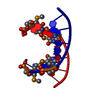
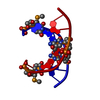
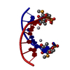
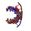
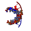
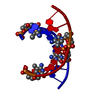
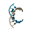
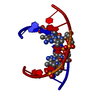
| Title | 5-SMe-dU containing DNA octamer | ||||||||||||||||||
 Components Components | 5'-D(* Keywords KeywordsDNA / Selenium / Nucleic Acid / 5-SMe-deoxyuridine / 2'-SeMe-deoxyuridine | Function / homology | DNA |  Function and homology information Function and homology informationMethod |  X-RAY DIFFRACTION / SYNCHROTRON / MOLECULAR REPLACEMENT / Resolution: 1.38 Å X-RAY DIFFRACTION / SYNCHROTRON / MOLECULAR REPLACEMENT / Resolution: 1.38 Å  Authors AuthorsSheng, J. / Hassan, A.E.A. / Zhang, W. / Gan, J. / Huang, Z. |  CitationJournal: Nucleic Acids Res. / Year: 2012 CitationJournal: Nucleic Acids Res. / Year: 2012Title: Hydrogen bond formation between the naturally modified nucleobase and phosphate backbone. Authors: Sheng, J. / Zhang, W. / Hassan, A.E. / Gan, J. / Soares, A.S. / Geng, S. / Ren, Y. / Huang, Z. History |
|
- Structure visualization
| Structure viewer | Molecule: MolmilJmol/JSmol |
|---|
- Downloads & links
-Download
| PDBx/mmCIF format | 3iki.cif.gz | 15.8 KB | Display | PDBx/mmCIF format |
|---|---|---|---|---|
| PDB format | pdb3iki.ent.gz | 9.6 KB | Display | PDB format |
| PDBx/mmJSON format | 3iki.json.gz | Tree view | PDBx/mmJSON format | |
| Others |  Other downloads Other downloads |
-Validation report
| Summary document | 3iki_validation.pdf.gz | 370.7 KB | Display | wwPDB validaton report |
|---|---|---|---|---|
| Full document | 3iki_full_validation.pdf.gz | 370.7 KB | Display | |
| Data in XML | 3iki_validation.xml.gz | 3 KB | Display | |
| Data in CIF | 3iki_validation.cif.gz | 3.5 KB | Display | |
| Arichive directory | https://data.pdbj.org/pub/pdb/validation_reports/ik/3ikiftp://data.pdbj.org/pub/pdb/validation_reports/ik/3iki | HTTPS FTP |
-Related structure data
| Related structure data | 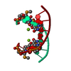 3ltrC  1dnsS S: Starting model for refinement C: citing same article ( |
|---|---|
| Similar structure data |

-Links
 PDBj
PDBj

- Assembly
| Deposited unit | 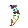
| ||||||||
|---|---|---|---|---|---|---|---|---|---|
| 1 | 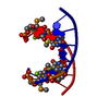
| ||||||||
| Unit cell |
|
-Components
| #1: DNA chain | Mass: 2537.642 Da / Num. of mol.: 1 / Source method: obtained synthetically / Details: Synthesized by solid phase synthesis |
|---|---|
| #2: Chemical | ChemComp-MG /   Mass: 24.305 Da / Num. of mol.: 1 / Source method: obtained synthetically / Formula: Mg Mass: 24.305 Da / Num. of mol.: 1 / Source method: obtained synthetically / Formula: Mg |
| #3: Water | ChemComp-HOH /  Mass: 18.015 Da / Num. of mol.: 38 / Source method: isolated from a natural source / Formula: H2O Mass: 18.015 Da / Num. of mol.: 38 / Source method: isolated from a natural source / Formula: H2O |
-Experimental details
-Experiment
| Experiment | Method: X-RAY DIFFRACTION / Number of used crystals: 1 |
|---|
- Sample preparation
| Crystal | Density Matthews: 2.15 Å3/Da / Density % sol: 42.67 % |
|---|---|
| Crystal grow | Temperature: 293 K / Method: vapor diffusion, hanging drop / pH: 7 Details: 10% v / v MPD, 40 mM Na Cacodylate pH 7.0, 12 mM Spermine tetra-HCl, 40 mM Lithium Chloride, 80 mM Strontium Chloride / 20 mM Magnesium Chloride, VAPOR DIFFUSION, HANGING DROP, temperature 293K |
-Data collection
| Diffraction | Mean temperature: 99 K |
|---|---|
| Diffraction source | Source: SYNCHROTRON / Site: NSLS  / Beamline: X12B / Wavelength: 0.9795 Å / Beamline: X12B / Wavelength: 0.9795 Å |
| Detector | Type: ADSC QUANTUM 4 / Detector: CCD / Date: Oct 22, 2008 |
| Radiation | Monochromator: channel-cut Si(111) monochromator / Protocol: SINGLE WAVELENGTH / Monochromatic (M) / Laue (L): M / Scattering type: x-ray |
| Radiation wavelength | Wavelength: 0.9795 Å / Relative weight: 1 |
| Reflection | Resolution: 1.38→50 Å / Num. all: 4911 / Num. obs: 4616 / % possible obs: 94 % / Redundancy: 8.4 % / Rmerge(I) obs: 0.057 / Net I/σ(I): 23.02 |
| Reflection shell | Resolution: 1.38→1.43 Å / Redundancy: 5.2 % / Rmerge(I) obs: 0.245 / Mean I/σ(I) obs: 5.86 / Num. unique all: 324 / % possible all: 67.8 |
- Processing
| Software |
| ||||||||||||||||||||||||||||||||||||||||||||||||||||||||||||
|---|---|---|---|---|---|---|---|---|---|---|---|---|---|---|---|---|---|---|---|---|---|---|---|---|---|---|---|---|---|---|---|---|---|---|---|---|---|---|---|---|---|---|---|---|---|---|---|---|---|---|---|---|---|---|---|---|---|---|---|---|---|
| Refinement | Method to determine structure: MOLECULAR REPLACEMENT Starting model: 1DNS Resolution: 1.38→30.32 Å / Cor.coef. Fo:Fc: 0.957 / Cor.coef. Fo:Fc free: 0.961 / SU B: 1.592 / SU ML: 0.031 / TLS residual ADP flag: LIKELY RESIDUAL / Cross valid method: THROUGHOUT / ESU R: 0.061 / ESU R Free: 0.06 / Stereochemistry target values: MAXIMUM LIKELIHOOD / Details: HYDROGENS HAVE BEEN ADDED IN THE RIDING POSITIONS
| ||||||||||||||||||||||||||||||||||||||||||||||||||||||||||||
| Solvent computation | Ion probe radii: 0.8 Å / Shrinkage radii: 0.8 Å / VDW probe radii: 1.2 Å / Solvent model: MASK | ||||||||||||||||||||||||||||||||||||||||||||||||||||||||||||
| Displacement parameters | Biso mean: 12.133 Å2
| ||||||||||||||||||||||||||||||||||||||||||||||||||||||||||||
| Refine analyze |
| ||||||||||||||||||||||||||||||||||||||||||||||||||||||||||||
| Refinement step | Cycle: LAST / Resolution: 1.38→30.32 Å
| ||||||||||||||||||||||||||||||||||||||||||||||||||||||||||||
| Refine LS restraints |
| ||||||||||||||||||||||||||||||||||||||||||||||||||||||||||||
| LS refinement shell | Resolution: 1.38→1.416 Å / Total num. of bins used: 20
| ||||||||||||||||||||||||||||||||||||||||||||||||||||||||||||
| Refinement TLS params. | Method: refined / Origin x: 31.5133 Å / Origin y: 30.4882 Å / Origin z: 2.3842 Å
|
