 Movie
Movie Controller

[English] 日本語
 Yorodumi
- PDB-3ijt: Structural Characterization of SMU.440, a Hypothetical Protein fr... -
+ Open data

- Basic information
| Entry | Database: PDB / ID: 3ijt | |||||||||
|---|---|---|---|---|---|---|---|---|---|---|
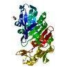
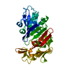
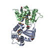
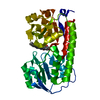
| Title | Structural Characterization of SMU.440, a Hypothetical Protein from Streptococcus mutans | |||||||||
 Components Components | Putative uncharacterized protein | |||||||||
 Keywords Keywords | UNKNOWN FUNCTION / HYPOTHETICAL PROTEIN | |||||||||
| Function / homology | START domain / Alpha-D-Glucose-1,6-Bisphosphate; Chain A, domain 4 / START-like domain superfamily / 2-Layer Sandwich / Alpha Beta / Polyketide cyclase Function and homology information Function and homology information | |||||||||
| Biological species |  Streptococcus mutans (bacteria) Streptococcus mutans (bacteria) | |||||||||
| Method |  X-RAY DIFFRACTION / SYNCHROTRON / SIRAS / Resolution: 2.377 Å X-RAY DIFFRACTION / SYNCHROTRON / SIRAS / Resolution: 2.377 Å | |||||||||
 Authors Authors | Nan, J. / Brostromer, E. / Kristensen, O. / Su, X.-D. | |||||||||
 Citation Citation | Journal: Plos One / Year: 2009 Title: Bioinformatics and structural characterization of a hypothetical protein from Streptococcus mutans: implication of antibiotic resistance Authors: Nan, J. / Brostromer, E. / Liu, X.-Y. / Kristensen, O. / Su, X.-D. | |||||||||
| History |
|
- Structure visualization
| Structure viewer | Molecule: MolmilJmol/JSmol |
|---|
- Downloads & links
-Download
| PDBx/mmCIF format | 3ijt.cif.gz | 69.3 KB | Display | PDBx/mmCIF format |
|---|---|---|---|---|
| PDB format | pdb3ijt.ent.gz | 52.2 KB | Display | PDB format |
| PDBx/mmJSON format | 3ijt.json.gz | Tree view | PDBx/mmJSON format | |
| Others |  Other downloads Other downloads |
-Validation report
| Arichive directory | https://data.pdbj.org/pub/pdb/validation_reports/ij/3ijtftp://data.pdbj.org/pub/pdb/validation_reports/ij/3ijt | HTTPS FTP |
|---|
-Related structure data
| Similar structure data |
|---|


-Links
 PDBj
PDBj- Assembly
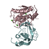
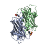
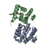
| Deposited unit | 
| ||||||||
|---|---|---|---|---|---|---|---|---|---|
| 1 |
| ||||||||
| Unit cell |
|
-Components
| #1: Protein | Mass: 17426.500 Da / Num. of mol.: 2 Source method: isolated from a genetically manipulated source Source: (gene. exp.) Streptococcus mutans (bacteria) / Strain: UA159 / Gene: SMU.440 / Plasmid: pET28A / Production host: #2: Water | ChemComp-HOH / |  Mass: 18.015 Da / Num. of mol.: 90 / Source method: isolated from a natural source / Formula: H2O Mass: 18.015 Da / Num. of mol.: 90 / Source method: isolated from a natural source / Formula: H2O |
|---|
-Experimental details
-Experiment
| Experiment | Method: X-RAY DIFFRACTION / Number of used crystals: 1 |
|---|
- Sample preparation
| Crystal |
| |||||||||||||||
|---|---|---|---|---|---|---|---|---|---|---|---|---|---|---|---|---|
| Crystal grow |
|
-Data collection
| Diffraction | Mean temperature: 100 K |
|---|---|
| Diffraction source | Source: SYNCHROTRON / Site: MAX II  / Beamline: I711 / Wavelength: 1.095 Å / Beamline: I711 / Wavelength: 1.095 Å |
| Detector | Type: MARRESEARCH / Detector: CCD / Date: Oct 4, 2004 |
| Radiation | Monochromator: asymmetrically cut Si(111) / Protocol: SINGLE WAVELENGTH / Monochromatic (M) / Laue (L): M / Scattering type: x-ray |
| Radiation wavelength | Wavelength: 1.095 Å / Relative weight: 1 |
| Reflection | Resolution: 2.377→34.078 Å / Num. obs: 17129 / % possible obs: 96.9 % / Observed criterion σ(F): 0 / Observed criterion σ(I): -3 / Redundancy: 2.7 % / Biso Wilson estimate: 50.51 Å2 / Rsym value: 0.038 / Net I/σ(I): 11.9 |
| Reflection shell | Resolution: 2.377→2.47 Å / Redundancy: 2.6 % / Mean I/σ(I) obs: 2.2 / Num. unique all: 1662 / Rsym value: 0.308 / % possible all: 97 |
- Processing
| Software |
| |||||||||||||||||||||||||||||||||||||||||||||||||
|---|---|---|---|---|---|---|---|---|---|---|---|---|---|---|---|---|---|---|---|---|---|---|---|---|---|---|---|---|---|---|---|---|---|---|---|---|---|---|---|---|---|---|---|---|---|---|---|---|---|---|
| Refinement | Method to determine structure: SIRAS / Resolution: 2.377→34.078 Å / Occupancy max: 1 / Occupancy min: 0 / FOM work R set: 0.791 / SU ML: 1.61 / Isotropic thermal model: Isotropic / σ(F): 0.07 / Phase error: 27.2 / Stereochemistry target values: ML
| |||||||||||||||||||||||||||||||||||||||||||||||||
| Solvent computation | Shrinkage radii: 0.9 Å / VDW probe radii: 1.11 Å / Solvent model: FLAT BULK SOLVENT MODEL / Bsol: 52.054 Å2 / ksol: 0.322 e/Å3 | |||||||||||||||||||||||||||||||||||||||||||||||||
| Displacement parameters | Biso max: 115.14 Å2 / Biso mean: 55.625 Å2 / Biso min: 33.01 Å2
| |||||||||||||||||||||||||||||||||||||||||||||||||
| Refinement step | Cycle: LAST / Resolution: 2.377→34.078 Å
| |||||||||||||||||||||||||||||||||||||||||||||||||
| Refine LS restraints |
| |||||||||||||||||||||||||||||||||||||||||||||||||
| LS refinement shell |
|
