 Movie
Movie Controller

[English] 日本語
 Yorodumi
- PDB-3igq: Crystal structure of the extracellular domain of a bacterial pent... -
+ Open data

- Basic information
| Entry | Database: PDB / ID: 3igq | ||||||
|---|---|---|---|---|---|---|---|
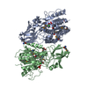
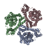
| Title | Crystal structure of the extracellular domain of a bacterial pentameric ligand-gated ion channel | ||||||
 Components Components | Glr4197 protein | ||||||
 Keywords Keywords | MEMBRANE PROTEIN / TRANSPORT PROTEIN / pLGIC cys-loop | ||||||
| Function / homology |  Function and homology information Function and homology informationsodium channel activity / potassium channel activity / extracellular ligand-gated monoatomic ion channel activity / transmembrane signaling receptor activity / identical protein binding / plasma membrane Similarity search - Function | ||||||
| Biological species |  Gloeobacter violaceus (bacteria) Gloeobacter violaceus (bacteria) | ||||||
| Method |  X-RAY DIFFRACTION / SYNCHROTRON / SIRAS / Resolution: 2.3 Å X-RAY DIFFRACTION / SYNCHROTRON / SIRAS / Resolution: 2.3 Å | ||||||
 Authors Authors | Nury, H. / Delarue, M. | ||||||
 Citation Citation | Journal: J.Mol.Biol. / Year: 2010 Title: Crystal structure of the extracellular domain of a bacterial ligand-gated ion channel Authors: Nury, H. / Bocquet, N. / Le Poupon, C. / Raynal, B. / Haouz, A. / Corringer, P.-J. / Delarue, M. | ||||||
| History |
|
- Structure visualization
| Structure viewer | Molecule: MolmilJmol/JSmol |
|---|
- Downloads & links
-Download
| PDBx/mmCIF format | 3igq.cif.gz | 231.5 KB | Display | PDBx/mmCIF format |
|---|---|---|---|---|
| PDB format | pdb3igq.ent.gz | 186.4 KB | Display | PDB format |
| PDBx/mmJSON format | 3igq.json.gz | Tree view | PDBx/mmJSON format | |
| Others |  Other downloads Other downloads |
-Validation report
| Arichive directory | https://data.pdbj.org/pub/pdb/validation_reports/ig/3igqftp://data.pdbj.org/pub/pdb/validation_reports/ig/3igq | HTTPS FTP |
|---|
-Related structure data
| Related structure data | |
|---|---|
| Similar structure data |


-Links
 PDBj
PDBj


- Assembly
| Deposited unit | 
| |||||||||||||||||||||
|---|---|---|---|---|---|---|---|---|---|---|---|---|---|---|---|---|---|---|---|---|---|---|
| 1 | 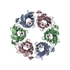
| |||||||||||||||||||||
| 2 | 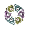
| |||||||||||||||||||||
| Unit cell |
| |||||||||||||||||||||
| Noncrystallographic symmetry (NCS) | NCS domain:
|
-Components
-Protein , 1 types, 6 molecules ABCDEF
| #1: Protein | Mass: 22872.697 Da / Num. of mol.: 6 Fragment: extracellular N-terminal fragment, UNP residues 44-235 Mutation: F116G, Y119T, P120E, F121S Source method: isolated from a genetically manipulated source Source: (gene. exp.) Gloeobacter violaceus (bacteria) / Gene: glr4197 / Plasmid: pET20b / Production host: |
|---|
-Non-polymers , 5 types, 432 molecules 


| #2: Chemical | ChemComp-ACY /  Mass: 60.052 Da / Num. of mol.: 6 / Source method: obtained synthetically / Formula: C2H4O2 Mass: 60.052 Da / Num. of mol.: 6 / Source method: obtained synthetically / Formula: C2H4O2#3: Chemical | ChemComp-NA /  Mass: 22.990 Da / Num. of mol.: 6 / Source method: obtained synthetically / Formula: Na Mass: 22.990 Da / Num. of mol.: 6 / Source method: obtained synthetically / Formula: Na#4: Chemical | ChemComp-HG /  Mass: 200.590 Da / Num. of mol.: 6 / Source method: obtained synthetically / Formula: Hg Mass: 200.590 Da / Num. of mol.: 6 / Source method: obtained synthetically / Formula: Hg#5: Chemical | ChemComp-CL /  Mass: 35.453 Da / Num. of mol.: 6 / Source method: obtained synthetically / Formula: Cl Mass: 35.453 Da / Num. of mol.: 6 / Source method: obtained synthetically / Formula: Cl#6: Water | ChemComp-HOH / | Mass: 18.015 Da / Num. of mol.: 408 / Source method: isolated from a natural source / Formula: H2O |
|---|
-Experimental details
-Experiment
| Experiment | Method: X-RAY DIFFRACTION / Number of used crystals: 1 |
|---|
- Sample preparation
| Crystal | Density Matthews: 2.28 Å3/Da / Density % sol: 45.96 % |
|---|
-Data collection
| Diffraction | Mean temperature: 100 K |
|---|---|
| Diffraction source | Source: SYNCHROTRON / Site: ESRF  / Beamline: ID14-1 / Wavelength: 0.934 Å / Beamline: ID14-1 / Wavelength: 0.934 Å |
| Detector | Type: ADSC QUANTUM 210 / Detector: CCD / Date: Aug 24, 2007 |
| Radiation | Protocol: SINGLE WAVELENGTH / Monochromatic (M) / Laue (L): M / Scattering type: x-ray |
| Radiation wavelength | Wavelength: 0.934 Å / Relative weight: 1 |
| Reflection | Resolution: 2.3→25 Å / Num. all: 108138 / Num. obs: 105975 / % possible obs: 98.4 % / Observed criterion σ(F): 0 / Observed criterion σ(I): -3 / Redundancy: 2.03 % / Rmerge(I) obs: 0.057 / Net I/σ(I): 11.23 |
| Reflection shell | Resolution: 2.3→2.44 Å / Redundancy: 2.01 % / Rmerge(I) obs: 0.424 / Mean I/σ(I) obs: 1.96 / % possible all: 97.91 |
- Processing
| Software |
| |||||||||||||||||||||||||||||||||||||||||||||||||||||||||||||||||||||||||||||||||||||||||||||||||||||||||||||||||||||||||||||
|---|---|---|---|---|---|---|---|---|---|---|---|---|---|---|---|---|---|---|---|---|---|---|---|---|---|---|---|---|---|---|---|---|---|---|---|---|---|---|---|---|---|---|---|---|---|---|---|---|---|---|---|---|---|---|---|---|---|---|---|---|---|---|---|---|---|---|---|---|---|---|---|---|---|---|---|---|---|---|---|---|---|---|---|---|---|---|---|---|---|---|---|---|---|---|---|---|---|---|---|---|---|---|---|---|---|---|---|---|---|---|---|---|---|---|---|---|---|---|---|---|---|---|---|---|---|---|
| Refinement | Method to determine structure: SIRAS / Resolution: 2.3→25 Å / Cor.coef. Fo:Fc: 0.942 / Cor.coef. Fo:Fc free: 0.913 / SU B: 7.36 / SU ML: 0.179 / Cross valid method: THROUGHOUT / ESU R: 0.353 / ESU R Free: 0.246 / Stereochemistry target values: MAXIMUM LIKELIHOOD Details: 1.HYDROGENS HAVE BEEN ADDED IN THE RIDING POSITIONS. 2.Friedel's pair are not merged during data collection (the anomalous information from the mercury ions were used), while the refinement ...Details: 1.HYDROGENS HAVE BEEN ADDED IN THE RIDING POSITIONS. 2.Friedel's pair are not merged during data collection (the anomalous information from the mercury ions were used), while the refinement is made with merged plus and minus reflections.
| |||||||||||||||||||||||||||||||||||||||||||||||||||||||||||||||||||||||||||||||||||||||||||||||||||||||||||||||||||||||||||||
| Solvent computation | Ion probe radii: 0.8 Å / Shrinkage radii: 0.8 Å / VDW probe radii: 1.4 Å / Solvent model: BABINET MODEL WITH MASK | |||||||||||||||||||||||||||||||||||||||||||||||||||||||||||||||||||||||||||||||||||||||||||||||||||||||||||||||||||||||||||||
| Displacement parameters | Biso mean: 50.431 Å2
| |||||||||||||||||||||||||||||||||||||||||||||||||||||||||||||||||||||||||||||||||||||||||||||||||||||||||||||||||||||||||||||
| Refinement step | Cycle: LAST / Resolution: 2.3→25 Å
| |||||||||||||||||||||||||||||||||||||||||||||||||||||||||||||||||||||||||||||||||||||||||||||||||||||||||||||||||||||||||||||
| Refine LS restraints |
| |||||||||||||||||||||||||||||||||||||||||||||||||||||||||||||||||||||||||||||||||||||||||||||||||||||||||||||||||||||||||||||
| Refine LS restraints NCS | Dom-ID: 1 / Ens-ID: 1 / Refine-ID: X-RAY DIFFRACTION
| |||||||||||||||||||||||||||||||||||||||||||||||||||||||||||||||||||||||||||||||||||||||||||||||||||||||||||||||||||||||||||||
| LS refinement shell | Resolution: 2.3→2.359 Å / Total num. of bins used: 20
|
