 Movie
Movie Controller


+ Open data

- Basic information
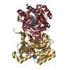
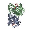
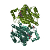
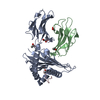
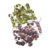
| Entry | Database: PDB / ID: 3if6 | ||||||
|---|---|---|---|---|---|---|---|
| Title | Crystal structure of OXA-46 beta-lactamase from P. aeruginosa | ||||||
 Components Components | (OXA-46 oxacillinase) x 3 | ||||||
 Keywords Keywords | HYDROLASE / serine beta-lactamase | ||||||
| Function / homology |  Function and homology information Function and homology informationpenicillin binding / antibiotic catabolic process / beta-lactamase activity / beta-lactamase / response to antibiotic Similarity search - Function | ||||||
| Biological species |   Pseudomonas aeruginosa (bacteria) Pseudomonas aeruginosa (bacteria) | ||||||
| Method |  X-RAY DIFFRACTION / SYNCHROTRON / MOLECULAR REPLACEMENT / Resolution: 2.4 Å X-RAY DIFFRACTION / SYNCHROTRON / MOLECULAR REPLACEMENT / Resolution: 2.4 Å | ||||||
 Authors Authors | Docquier, J.D. / Benvenuti, M. / Calderone, V. / Giuliani, F. / Kapetis, D. / De Luca, F. / Rossolini, G.M. / Mangani, S. | ||||||
 Citation Citation | Journal: Antimicrob.Agents Chemother. / Year: 2010 Title: Crystal structure of the narrow-spectrum OXA-46 class D beta-lactamase: relationship between active-site lysine carbamylation and inhibition by polycarboxylates Authors: Docquier, J.D. / Benvenuti, M. / Calderone, V. / Giuliani, F. / Kapetis, D. / De Luca, F. / Rossolini, G.M. / Mangani, S. #1: Journal: Antimicrob.Agents Chemother. / Year: 2005 Title: OXA-46, a new class D beta-lactamase of narrow substrate specificity encoded by a blaVIM-1-containing integron from a Pseudomonas aeruginosa clinical isolate Authors: Giuliani, F. / Docquier, J.D. / Riccio, M.L. / Pagani, L. / Rossolini, G.M. | ||||||
| History |
|
- Structure visualization
| Structure viewer | Molecule: MolmilJmol/JSmol |
|---|
- Downloads & links
-Download
| PDBx/mmCIF format | 3if6.cif.gz | 167.9 KB | Display | PDBx/mmCIF format |
|---|---|---|---|---|
| PDB format | pdb3if6.ent.gz | 132 KB | Display | PDB format |
| PDBx/mmJSON format | 3if6.json.gz | Tree view | PDBx/mmJSON format | |
| Others |  Other downloads Other downloads |
-Validation report
| Summary document | 3if6_validation.pdf.gz | 703.1 KB | Display | wwPDB validaton report |
|---|---|---|---|---|
| Full document | 3if6_full_validation.pdf.gz | 725.5 KB | Display | |
| Data in XML | 3if6_validation.xml.gz | 35.4 KB | Display | |
| Data in CIF | 3if6_validation.cif.gz | 49.1 KB | Display | |
| Arichive directory | https://data.pdbj.org/pub/pdb/validation_reports/if/3if6ftp://data.pdbj.org/pub/pdb/validation_reports/if/3if6 | HTTPS FTP |
-Related structure data
| Related structure data |  3hbrS S: Starting model for refinement |
|---|---|
| Similar structure data |


-Links
 PDBj
PDBj
- Assembly
| Deposited unit | 
| ||||||||
|---|---|---|---|---|---|---|---|---|---|
| 1 | 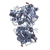
| ||||||||
| 2 | 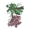
| ||||||||
| Unit cell |
|
-Components
-Protein , 3 types, 3 molecules ABC
| #1: Protein | Mass: 31140.367 Da / Num. of mol.: 1 Source method: isolated from a genetically manipulated source Source: (gene. exp.) Pseudomonas aeruginosa (bacteria) / Strain: PPV-97 / Plasmid: peT-9a / Production host: |
|---|---|
| #2: Protein | Mass: 31097.367 Da / Num. of mol.: 1 Source method: isolated from a genetically manipulated source Source: (gene. exp.) Pseudomonas aeruginosa (bacteria) / Strain: PPV-97 / Plasmid: peT-9a / Production host: |
| #3: Protein | Mass: 31140.367 Da / Num. of mol.: 1 Source method: isolated from a genetically manipulated source Source: (gene. exp.) Pseudomonas aeruginosa (bacteria) / Strain: PPV-97 / Plasmid: peT-9a / Production host: |
-Non-polymers , 4 types, 407 molecules 


| #4: Chemical |  Mass: 150.087 Da / Num. of mol.: 2 / Source method: obtained synthetically / Formula: C4H6O6 Mass: 150.087 Da / Num. of mol.: 2 / Source method: obtained synthetically / Formula: C4H6O6#5: Chemical | ChemComp-EDO /  Mass: 62.068 Da / Num. of mol.: 6 / Source method: obtained synthetically / Formula: C2H6O2 Mass: 62.068 Da / Num. of mol.: 6 / Source method: obtained synthetically / Formula: C2H6O2#6: Chemical | ChemComp-P6G / |  Mass: 282.331 Da / Num. of mol.: 1 / Source method: obtained synthetically / Formula: C12H26O7 / Comment: precipitant*YM Mass: 282.331 Da / Num. of mol.: 1 / Source method: obtained synthetically / Formula: C12H26O7 / Comment: precipitant*YM#7: Water | ChemComp-HOH / | Mass: 18.015 Da / Num. of mol.: 398 / Source method: isolated from a natural source / Formula: H2O |
|---|
-Experimental details
-Experiment
| Experiment | Method: X-RAY DIFFRACTION / Number of used crystals: 1 |
|---|
- Sample preparation
| Crystal | Density Matthews: 2.59 Å3/Da / Density % sol: 52.53 % |
|---|---|
| Crystal grow | Temperature: 293 K / Method: vapor diffusion, sitting drop / pH: 7.5 Details: 1M Na,K L-tartrate tetrahydrate, 50mM Hepes, 2-4%(v/v) PEG 400, pH 7.5, VAPOR DIFFUSION, SITTING DROP, temperature 293K |
-Data collection
| Diffraction | Mean temperature: 100 K |
|---|---|
| Diffraction source | Source: SYNCHROTRON / Site: EMBL/DESY, HAMBURG  / Beamline: BW7A / Wavelength: 0.92 Å / Beamline: BW7A / Wavelength: 0.92 Å |
| Detector | Type: MAR CCD 165 mm / Detector: CCD / Date: Apr 28, 2005 |
| Radiation | Monochromator: Double crystal Si(111) / Protocol: SINGLE WAVELENGTH / Monochromatic (M) / Laue (L): M / Scattering type: x-ray |
| Radiation wavelength | Wavelength: 0.92 Å / Relative weight: 1 |
| Reflection | Resolution: 2.2→56.5 Å / Num. all: 48568 / Num. obs: 48227 / % possible obs: 98.7 % / Observed criterion σ(F): 0 / Observed criterion σ(I): 1.2 / Redundancy: 8.8 % / Biso Wilson estimate: 42.418 Å2 / Rmerge(I) obs: 0.065 / Rsym value: 0.065 / Net I/σ(I): 9.6 |
| Reflection shell | Resolution: 2.2→2.32 Å / Redundancy: 7.78 % / Rmerge(I) obs: 0.61 / Mean I/σ(I) obs: 1.2 / Num. unique all: 756 / Rsym value: 0.61 / % possible all: 98.7 |
- Processing
| Software |
| ||||||||||||||||||||||||||||||||||||||||||||||||||||||||||||||||||||||||||||||||||||||||||
|---|---|---|---|---|---|---|---|---|---|---|---|---|---|---|---|---|---|---|---|---|---|---|---|---|---|---|---|---|---|---|---|---|---|---|---|---|---|---|---|---|---|---|---|---|---|---|---|---|---|---|---|---|---|---|---|---|---|---|---|---|---|---|---|---|---|---|---|---|---|---|---|---|---|---|---|---|---|---|---|---|---|---|---|---|---|---|---|---|---|---|---|
| Refinement | Method to determine structure: MOLECULAR REPLACEMENT Starting model: 3HBR Resolution: 2.4→31.4 Å / Cor.coef. Fo:Fc: 0.94 / Cor.coef. Fo:Fc free: 0.892 / SU B: 9.322 / SU ML: 0.219 / Cross valid method: THROUGHOUT / σ(F): 0 / σ(I): 2 / ESU R: 0.438 / ESU R Free: 0.303 / Stereochemistry target values: MAXIMUM LIKELIHOOD
| ||||||||||||||||||||||||||||||||||||||||||||||||||||||||||||||||||||||||||||||||||||||||||
| Solvent computation | Ion probe radii: 0.8 Å / Shrinkage radii: 0.8 Å / VDW probe radii: 1.2 Å / Solvent model: MASK | ||||||||||||||||||||||||||||||||||||||||||||||||||||||||||||||||||||||||||||||||||||||||||
| Displacement parameters | Biso mean: 40.818 Å2
| ||||||||||||||||||||||||||||||||||||||||||||||||||||||||||||||||||||||||||||||||||||||||||
| Refine analyze |
| ||||||||||||||||||||||||||||||||||||||||||||||||||||||||||||||||||||||||||||||||||||||||||
| Refinement step | Cycle: LAST / Resolution: 2.4→31.4 Å
| ||||||||||||||||||||||||||||||||||||||||||||||||||||||||||||||||||||||||||||||||||||||||||
| Refine LS restraints |
| ||||||||||||||||||||||||||||||||||||||||||||||||||||||||||||||||||||||||||||||||||||||||||
| LS refinement shell | Resolution: 2.4→2.462 Å / Total num. of bins used: 20
|
