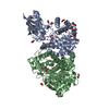
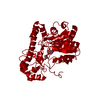
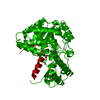
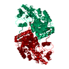
ANALYTICAL SIZE EXCLUSION CHROMATOGRAPHY WITH STATIC LIGHT SCATTERING SUPPORTS THE ASSIGNMENT OF A DIMER AS A SIGNIFICANT OLIGOMERIZATION STATE IN SOLUTION.
-
Components
#1: Protein
Aminotransferase
Mass: 49601.477 Da / Num. of mol.: 2 Source method: isolated from a genetically manipulated source Source: (gene. exp.) Psychrobacter arcticus 273-4 (bacteria) Strain: DSM 17307 / 273-4 / Gene: avtA, Psyc_2118, YP_265399.1 / Plasmid: SpeedET / Production host: Escherichia coli (E. coli) / Strain (production host): HK100 References: UniProt: Q4FPU3, Transferases; Transferring nitrogenous groups; Transaminases
Mass: 18.015 Da / Num. of mol.: 203 / Source method: isolated from a natural source / Formula: H2O
Sequence details
SEQUENCE THIS CONSTRUCT WAS EXPRESSED WITH A PURIFICATION TAG MGSDKIHHHHHHENLYFQG. THE TAG WAS ...SEQUENCE THIS CONSTRUCT WAS EXPRESSED WITH A PURIFICATION TAG MGSDKIHHHHHHENLYFQG. THE TAG WAS REMOVED WITH TEV PROTEASE LEAVING ONLY A GLYCINE (0) FOLLOWED BY THE TARGET SEQUENCE.
-
Experimental details
-
Experiment
Experiment
Method: X-RAY DIFFRACTION / Number of used crystals: 1
-
Sample preparation
Crystal
Density Matthews: 2.96 Å3/Da / Density % sol: 58.45 % Description: DATA WERE SCALED USING XSCALE WITH FRIEDEL PAIRS KEPT AS SEPARATE WHEN COMPUTING R-SYM, COMPLETENESS AND .
Crystal grow
Temperature: 293 K / Method: vapor diffusion, sitting drop / pH: 5.6 Details: 0.1700M ammonium acetate, 15.0000% Glycerol, 25.5000% polyethylene glycol 4000, 0.1M sodium citrate pH 5.6, Additive: 0.001 M FeCl2, NANODROP, VAPOR DIFFUSION, SITTING DROP, temperature 293K
Resolution: 2.5→29.975 Å / Num. obs: 39646 / % possible obs: 93.7 % / Observed criterion σ(I): -3 / Redundancy: 3.79 % / Biso Wilson estimate: 47.923 Å2 / Rmerge(I) obs: 0.075 / Net I/σ(I): 9.14
Reflection shell
Resolution (Å)
Rmerge(I) obs
Mean I/σ(I) obs
Num. measured obs
Num. unique obs
Diffraction-ID
% possible all
2.5-2.59
0.438
2
13620
6839
1
85.1
2.59-2.69
0.369
2.5
14491
7146
1
93.5
2.69-2.81
0.284
3.2
14932
7354
1
94.5
2.81-2.96
0.216
4.1
15496
7643
1
94.7
2.96-3.15
0.149
5.7
15671
7727
1
94.6
3.15-3.39
0.106
7.7
15114
7461
1
95.2
3.39-3.73
0.068
11.2
15030
7445
1
94.1
3.73-4.26
0.043
15.6
15069
7471
1
95.1
4.26-5.35
0.04
18.2
15164
7510
1
94.9
5.35-29.975
0.035
20.1
15552
7709
1
95.1
-
Phasing
Phasing
Method: MAD
-
Processing
Software
Name
Version
Classification
NB
REFMAC
5.5.0053
refinement
PHENIX
refinement
SHELX
phasing
MolProbity
3beta29
modelbuilding
XSCALE
datascaling
PDB_EXTRACT
3.006
dataextraction
XDS
datareduction
SHELXD
phasing
autoSHARP
phasing
Refinement
Method to determine structure: MAD / Resolution: 2.5→29.975 Å / Cor.coef. Fo:Fc: 0.952 / Cor.coef. Fo:Fc free: 0.926 / Occupancy max: 1 / Occupancy min: 0.4 / SU B: 17.234 / SU ML: 0.171 / TLS residual ADP flag: LIKELY RESIDUAL / Cross valid method: THROUGHOUT / σ(F): 0 / ESU R: 0.4 / ESU R Free: 0.245 Stereochemistry target values: MAXIMUM LIKELIHOOD WITH PHASES Details: 1. HYDROGENS HAVE BEEN ADDED IN THE RIDING POSITIONS. 2. ATOM RECORDS CONTAIN RESIDUAL B FACTORS ONLY. 3. A MET-INHIBITION PROTOCOL WAS USED FOR SELENOMETHIONINE INCORPORATION DURING PROTEIN ...Details: 1. HYDROGENS HAVE BEEN ADDED IN THE RIDING POSITIONS. 2. ATOM RECORDS CONTAIN RESIDUAL B FACTORS ONLY. 3. A MET-INHIBITION PROTOCOL WAS USED FOR SELENOMETHIONINE INCORPORATION DURING PROTEIN EXPRESSION. THE OCCUPANCY OF THE SE ATOMS IN THE MSE RESIDUES WAS REDUCED TO 0.75 FOR THE REDUCED SCATTERING POWER DUE TO PARTIAL S-MET INCORPORATION. 4. GLYCEROL (GOL) AND ACETATE (ACT) MOLECULES FROM THE CRYSTALLIZATION SOLUTION ARE MODELED. 5. THE LIGAND PYRIDOXAL-5'-PHOSPHATE (PLP) IS MODELED INTO BOTH MONOMERS.
Rfactor
Num. reflection
% reflection
Selection details
Rfree
0.221
1986
5 %
RANDOM
Rwork
0.178
-
-
-
obs
0.181
39646
98.82 %
-
Solvent computation
Ion probe radii: 0.8 Å / Shrinkage radii: 0.8 Å / VDW probe radii: 1.4 Å / Solvent model: BABINET MODEL WITH MASK
In the structure databanks used in Yorodumi, some data are registered as the other names, "COVID-19 virus" and "2019-nCoV". Here are the details of the virus and the list of structure data.
Jan 31, 2019. EMDB accession codes are about to change! (news from PDBe EMDB page)
EMDB accession codes are about to change! (news from PDBe EMDB page)
The allocation of 4 digits for EMDB accession codes will soon come to an end. Whilst these codes will remain in use, new EMDB accession codes will include an additional digit and will expand incrementally as the available range of codes is exhausted. The current 4-digit format prefixed with “EMD-” (i.e. EMD-XXXX) will advance to a 5-digit format (i.e. EMD-XXXXX), and so on. It is currently estimated that the 4-digit codes will be depleted around Spring 2019, at which point the 5-digit format will come into force.
The EM Navigator/Yorodumi systems omit the EMD- prefix.
Related info.:Q: What is EMD? / ID/Accession-code notation in Yorodumi/EM Navigator
Yorodumi is a browser for structure data from EMDB, PDB, SASBDB, etc.
This page is also the successor to EM Navigator detail page, and also detail information page/front-end page for Omokage search.
The word "yorodu" (or yorozu) is an old Japanese word meaning "ten thousand". "mi" (miru) is to see.
Related info.:EMDB / PDB / SASBDB / Comparison of 3 databanks / Yorodumi Search / Aug 31, 2016. New EM Navigator & Yorodumi / Yorodumi Papers / Jmol/JSmol / Function and homology information / Changes in new EM Navigator and Yorodumi
 Movie
Movie Controller

 Yorodumi
 Open data
 Basic information
 Components
 Keywords
 Function and homology information
 Psychrobacter arcticus 273-4 (bacteria)
 X-RAY DIFFRACTION /
  Authors
 Citation
 Structure visualization
 Downloads & links
 Other downloads

 PDBj
PDBj Assembly

 Mass: 247.142 Da / Num. of mol.: 2 / Source method: obtained synthetically / Formula: C8H10NO6P
 Mass: 92.094 Da / Num. of mol.: 2 / Source method: obtained synthetically / Formula: C3H8O3
 Mass: 59.044 Da / Num. of mol.: 1 / Source method: obtained synthetically / Formula: C2H3O2
 Mass: 18.015 Da / Num. of mol.: 203 / Source method: isolated from a natural source / Formula: H2O
 Sample preparation
 / Beamline: BL9-2 / Wavelength: 0.91162,0.97934,0.97920
 Processing