 Movie
Movie Controller


+ Open data

- Basic information
| Entry | Database: PDB / ID: 3i85 | ||||||
|---|---|---|---|---|---|---|---|
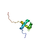
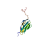
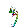
| Title | The Crystal Structure of Human EMMPRIN N-terminal Domain 1 | ||||||
 Components Components | Cervical EMMPRIN | ||||||
 Keywords Keywords | CELL ADHESION / EMMPRIN / CD147 / dimerization / beta-strand swapping | ||||||
| Function / homology |  Function and homology information Function and homology informationDefective SLC16A1 causes symptomatic deficiency in lactate transport (SDLT) / Proton-coupled monocarboxylate transport / positive regulation of matrix metallopeptidase secretion / acrosomal membrane / dendrite self-avoidance / endothelial tube morphogenesis / response to mercury ion / cell-cell adhesion mediator activity / neural retina development / photoreceptor cell maintenance ...Defective SLC16A1 causes symptomatic deficiency in lactate transport (SDLT) / Proton-coupled monocarboxylate transport / positive regulation of matrix metallopeptidase secretion / acrosomal membrane / dendrite self-avoidance / endothelial tube morphogenesis / response to mercury ion / cell-cell adhesion mediator activity / neural retina development / photoreceptor cell maintenance / neuron projection extension / Basigin interactions / Aspirin ADME / odontogenesis of dentin-containing tooth / D-mannose binding / homophilic cell-cell adhesion / decidualization / positive regulation of vascular endothelial growth factor production / Integrin cell surface interactions / photoreceptor outer segment / response to cAMP / Degradation of the extracellular matrix / neutrophil chemotaxis / photoreceptor inner segment / embryo implantation / positive regulation of endothelial cell migration / axon guidance / protein localization to plasma membrane / sarcolemma / positive regulation of interleukin-6 production / response to peptide hormone / melanosome / virus receptor activity / signaling receptor activity / angiogenesis / basolateral plasma membrane / positive regulation of viral entry into host cell / cell surface receptor signaling pathway / endosome / cadherin binding / Golgi membrane / axon / focal adhesion / endoplasmic reticulum membrane / mitochondrion / extracellular exosome / membrane / plasma membrane Similarity search - Function | ||||||
| Biological species |  Homo sapiens (human) Homo sapiens (human) | ||||||
| Method |  X-RAY DIFFRACTION / MOLECULAR REPLACEMENT / Resolution: 2.5 Å X-RAY DIFFRACTION / MOLECULAR REPLACEMENT / Resolution: 2.5 Å | ||||||
 Authors Authors | Luo, J. / Gilliland, G.L. | ||||||
 Citation Citation | Journal: Proteins / Year: 2009 Title: Structure of the EMMPRIN N-terminal domain 1: Dimerization via beta-strand swapping. Authors: Luo, J. / Teplyakov, A. / Obmolova, G. / Malia, T. / Wu, S.J. / Beil, E. / Baker, A. / Swencki-Underwood, B. / Zhao, Y. / Sprenkle, J. / Dixon, K. / Sweet, R. / Gilliland, G.L. | ||||||
| History |
|
- Structure visualization
| Structure viewer | Molecule: MolmilJmol/JSmol |
|---|
- Downloads & links
-Download
| PDBx/mmCIF format | 3i85.cif.gz | 77.5 KB | Display | PDBx/mmCIF format |
|---|---|---|---|---|
| PDB format | pdb3i85.ent.gz | 58.3 KB | Display | PDB format |
| PDBx/mmJSON format | 3i85.json.gz | Tree view | PDBx/mmJSON format | |
| Others |  Other downloads Other downloads |
-Validation report
| Arichive directory | https://data.pdbj.org/pub/pdb/validation_reports/i8/3i85ftp://data.pdbj.org/pub/pdb/validation_reports/i8/3i85 | HTTPS FTP |
|---|
-Related structure data
| Related structure data | 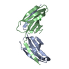 3i84C 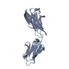 3b5hS C: citing same article ( S: Starting model for refinement |
|---|---|
| Similar structure data |


-Links
 PDBj
PDBj


- Assembly
| Deposited unit | 
| |||||||||||||||||||||||||||||||||||||||||||||||||
|---|---|---|---|---|---|---|---|---|---|---|---|---|---|---|---|---|---|---|---|---|---|---|---|---|---|---|---|---|---|---|---|---|---|---|---|---|---|---|---|---|---|---|---|---|---|---|---|---|---|---|
| 1 | 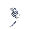
| |||||||||||||||||||||||||||||||||||||||||||||||||
| 2 | 
| |||||||||||||||||||||||||||||||||||||||||||||||||
| Unit cell |
| |||||||||||||||||||||||||||||||||||||||||||||||||
| Components on special symmetry positions |
| |||||||||||||||||||||||||||||||||||||||||||||||||
| Noncrystallographic symmetry (NCS) | NCS domain:
NCS domain segments:
NCS ensembles :
NCS oper:
|
-Components
| #1: Protein | Mass: 10558.675 Da / Num. of mol.: 2 / Mutation: A19D, N44D Source method: isolated from a genetically manipulated source Source: (gene. exp.) Homo sapiens (human) / Gene: hEMMPRIN, BSG, hCG_20562 / Plasmid: pCEP4 / Cell line (production host): HEK293 / Organ (production host): KIDNEY / Production host: Homo Sapiens (human) / References: UniProt: Q54A51, UniProt: P35613*PLUS#2: Water | ChemComp-HOH / |  Mass: 18.015 Da / Num. of mol.: 12 / Source method: isolated from a natural source / Formula: H2O Mass: 18.015 Da / Num. of mol.: 12 / Source method: isolated from a natural source / Formula: H2OHas protein modification | Y | |
|---|
-Experimental details
-Experiment
| Experiment | Method: X-RAY DIFFRACTION / Number of used crystals: 1 |
|---|
- Sample preparation
| Crystal | Density Matthews: 2.4 Å3/Da / Density % sol: 48.74 % |
|---|---|
| Crystal grow | Temperature: 298 K / Method: vapor diffusion, sitting drop / pH: 6.5 Details: 3.2 M sodium formate, 3% MPD, 0.1 M sodium citrate, pH 3.5, VAPOR DIFFUSION, SITTING DROP, temperature 298K |
-Data collection
| Diffraction | Mean temperature: 118 K |
|---|---|
| Diffraction source | Source: ROTATING ANODE / Type: RIGAKU MICROMAX-007 HF / Wavelength: 1.5418 Å |
| Detector | Type: RIGAKU SATURN 944 / Detector: CCD / Date: Jun 7, 2007 / Details: Osmic VariMax Confocal |
| Radiation | Monochromator: Osmic VariMax Confocal / Protocol: SINGLE WAVELENGTH / Monochromatic (M) / Laue (L): M / Scattering type: x-ray |
| Radiation wavelength | Wavelength: 1.5418 Å / Relative weight: 1 |
| Reflection | Resolution: 2.5→42 Å / Num. all: 7149 / Num. obs: 7149 / % possible obs: 100 % / Observed criterion σ(F): 0 / Observed criterion σ(I): -3 / Redundancy: 18.4 % / Biso Wilson estimate: 95.5 Å2 / Rmerge(I) obs: 0.087 / Rsym value: 0.087 / Net I/σ(I): 17.1 |
| Reflection shell | Resolution: 2.5→2.59 Å / Redundancy: 10.8 % / Rmerge(I) obs: 0.591 / Mean I/σ(I) obs: 2.6 / Rsym value: 0.591 / % possible all: 100 |
- Processing
| Software |
| ||||||||||||||||||||||||||||||||||||||||||||||||||||||||||||||||||||||||||||||||||||||||||||||||||||||||||||||||||||||||||||||||||||||||||||||||||||||||||||
|---|---|---|---|---|---|---|---|---|---|---|---|---|---|---|---|---|---|---|---|---|---|---|---|---|---|---|---|---|---|---|---|---|---|---|---|---|---|---|---|---|---|---|---|---|---|---|---|---|---|---|---|---|---|---|---|---|---|---|---|---|---|---|---|---|---|---|---|---|---|---|---|---|---|---|---|---|---|---|---|---|---|---|---|---|---|---|---|---|---|---|---|---|---|---|---|---|---|---|---|---|---|---|---|---|---|---|---|---|---|---|---|---|---|---|---|---|---|---|---|---|---|---|---|---|---|---|---|---|---|---|---|---|---|---|---|---|---|---|---|---|---|---|---|---|---|---|---|---|---|---|---|---|---|---|---|---|---|
| Refinement | Method to determine structure: MOLECULAR REPLACEMENT Starting model: PDB CODE 3B5H Resolution: 2.5→29.237 Å / Occupancy max: 1 / Occupancy min: 1 / SU ML: 0.98 / σ(F): 1.34 / Phase error: 35.52 / Stereochemistry target values: ML
| ||||||||||||||||||||||||||||||||||||||||||||||||||||||||||||||||||||||||||||||||||||||||||||||||||||||||||||||||||||||||||||||||||||||||||||||||||||||||||||
| Solvent computation | Shrinkage radii: 0.9 Å / VDW probe radii: 1.11 Å / Solvent model: FLAT BULK SOLVENT MODEL / Bsol: 95.124 Å2 / ksol: 0.38 e/Å3 | ||||||||||||||||||||||||||||||||||||||||||||||||||||||||||||||||||||||||||||||||||||||||||||||||||||||||||||||||||||||||||||||||||||||||||||||||||||||||||||
| Displacement parameters | Biso mean: 90.571 Å2
| ||||||||||||||||||||||||||||||||||||||||||||||||||||||||||||||||||||||||||||||||||||||||||||||||||||||||||||||||||||||||||||||||||||||||||||||||||||||||||||
| Refinement step | Cycle: LAST / Resolution: 2.5→29.237 Å
| ||||||||||||||||||||||||||||||||||||||||||||||||||||||||||||||||||||||||||||||||||||||||||||||||||||||||||||||||||||||||||||||||||||||||||||||||||||||||||||
| Refine LS restraints |
| ||||||||||||||||||||||||||||||||||||||||||||||||||||||||||||||||||||||||||||||||||||||||||||||||||||||||||||||||||||||||||||||||||||||||||||||||||||||||||||
| Refine LS restraints NCS |
| ||||||||||||||||||||||||||||||||||||||||||||||||||||||||||||||||||||||||||||||||||||||||||||||||||||||||||||||||||||||||||||||||||||||||||||||||||||||||||||
| LS refinement shell |
| ||||||||||||||||||||||||||||||||||||||||||||||||||||||||||||||||||||||||||||||||||||||||||||||||||||||||||||||||||||||||||||||||||||||||||||||||||||||||||||
| Refinement TLS params. | Refine-ID: X-RAY DIFFRACTION
| ||||||||||||||||||||||||||||||||||||||||||||||||||||||||||||||||||||||||||||||||||||||||||||||||||||||||||||||||||||||||||||||||||||||||||||||||||||||||||||
| Refinement TLS group | Selection details: chain B and resid 93:103 |
