 Movie
Movie Controller

[English] 日本語
 Yorodumi
- PDB-3i4o: Crystal Structure of Translation Initiation Factor 1 from Mycobac... -
+ Open data

- Basic information
| Entry | Database: PDB / ID: 3i4o | ||||||
|---|---|---|---|---|---|---|---|
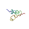
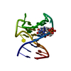
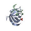
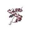
| Title | Crystal Structure of Translation Initiation Factor 1 from Mycobacterium tuberculosis | ||||||
 Components Components | Translation initiation factor IF-1 | ||||||
 Keywords Keywords | TRANSLATION / translation initiation / IF1 / Initiation factor / Protein biosynthesis | ||||||
| Function / homology |  Function and homology information Function and homology informationtranslation initiation factor activity / peptidoglycan-based cell wall / ribosome binding / rRNA binding / plasma membrane / cytoplasm / cytosol Similarity search - Function | ||||||
| Biological species |   Mycobacterium tuberculosis (bacteria) Mycobacterium tuberculosis (bacteria) | ||||||
| Method |  X-RAY DIFFRACTION / SYNCHROTRON / SAD / Resolution: 1.47 Å X-RAY DIFFRACTION / SYNCHROTRON / SAD / Resolution: 1.47 Å | ||||||
 Authors Authors | Hatzopoulos, G.N. / Mueller-Dieckmann, J. | ||||||
 Citation Citation | Journal: Febs Lett. / Year: 2010 Title: Structure of translation initiation factor 1 from Mycobacterium tuberculosis and inferred binding to the 30S ribosomal subunit. Authors: Hatzopoulos, G.N. / Mueller-Dieckmann, J. #1: Journal: Acta Crystallogr.,Sect.F / Year: 2007 Title: Cloning, expression, purification, crystallization and preliminary X-ray crystallographic analysis of initiation factor 1 from Mycobacterium tuberculosis. Authors: Hatzopoulos, G.N. / Mueller-Dieckmann, J. | ||||||
| History |
|
- Structure visualization
| Structure viewer | Molecule: MolmilJmol/JSmol |
|---|
- Downloads & links
-Download
| PDBx/mmCIF format | 3i4o.cif.gz | 76.1 KB | Display | PDBx/mmCIF format |
|---|---|---|---|---|
| PDB format | pdb3i4o.ent.gz | 57.9 KB | Display | PDB format |
| PDBx/mmJSON format | 3i4o.json.gz | Tree view | PDBx/mmJSON format | |
| Others |  Other downloads Other downloads |
-Validation report
| Arichive directory | https://data.pdbj.org/pub/pdb/validation_reports/i4/3i4oftp://data.pdbj.org/pub/pdb/validation_reports/i4/3i4o | HTTPS FTP |
|---|
-Related structure data
| Similar structure data |
|---|


-Links
 PDBj
PDBj
- Assembly
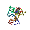
| Deposited unit | 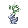
| ||||||||
|---|---|---|---|---|---|---|---|---|---|
| 1 | 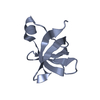
| ||||||||
| 2 | 
| ||||||||
| Unit cell |
|
-Components
| #1: Protein | Mass: 9135.647 Da / Num. of mol.: 2 Source method: isolated from a genetically manipulated source Source: (gene. exp.) Mycobacterium tuberculosis (bacteria) / Strain: H37RvGene: infA, MCB1222.32c, MT3568, MTCY13E12.15c, Rv3462c, Rv3462c (infA) Plasmid: pET151/D/TOPO / Production host: #2: Water | ChemComp-HOH / |  Mass: 18.015 Da / Num. of mol.: 163 / Source method: isolated from a natural source / Formula: H2O Mass: 18.015 Da / Num. of mol.: 163 / Source method: isolated from a natural source / Formula: H2O |
|---|
-Experimental details
-Experiment
| Experiment | Method: X-RAY DIFFRACTION / Number of used crystals: 2 |
|---|
- Sample preparation
| Crystal | Density Matthews: 2.17 Å3/Da / Density % sol: 43.34 % / Mosaicity: 0.5 ° |
|---|---|
| Crystal grow | Temperature: 292 K / Method: vapor diffusion, hanging drop / pH: 4.5 Details: 12-16% (w/v) PEG 8000, 50-200mM KCl and 8-16% (v/v) glycerol, pH 4.5, vapor diffusion, hanging drop, temperature 292K |
-Data collection
| Diffraction |
| |||||||||||||||||||||||||||||||||||||||||||||||||||||||||||||||||||||||||||||||||||||||||||||||||||||||||||||||||||||||||||||||||||||||||||||||||||
|---|---|---|---|---|---|---|---|---|---|---|---|---|---|---|---|---|---|---|---|---|---|---|---|---|---|---|---|---|---|---|---|---|---|---|---|---|---|---|---|---|---|---|---|---|---|---|---|---|---|---|---|---|---|---|---|---|---|---|---|---|---|---|---|---|---|---|---|---|---|---|---|---|---|---|---|---|---|---|---|---|---|---|---|---|---|---|---|---|---|---|---|---|---|---|---|---|---|---|---|---|---|---|---|---|---|---|---|---|---|---|---|---|---|---|---|---|---|---|---|---|---|---|---|---|---|---|---|---|---|---|---|---|---|---|---|---|---|---|---|---|---|---|---|---|---|---|---|---|
| Diffraction source |
| |||||||||||||||||||||||||||||||||||||||||||||||||||||||||||||||||||||||||||||||||||||||||||||||||||||||||||||||||||||||||||||||||||||||||||||||||||
| Detector |
| |||||||||||||||||||||||||||||||||||||||||||||||||||||||||||||||||||||||||||||||||||||||||||||||||||||||||||||||||||||||||||||||||||||||||||||||||||
| Radiation | Monochromator: Si [111], horizontally focussing / Protocol: SAD / Scattering type: x-ray | |||||||||||||||||||||||||||||||||||||||||||||||||||||||||||||||||||||||||||||||||||||||||||||||||||||||||||||||||||||||||||||||||||||||||||||||||||
| Radiation wavelength |
| |||||||||||||||||||||||||||||||||||||||||||||||||||||||||||||||||||||||||||||||||||||||||||||||||||||||||||||||||||||||||||||||||||||||||||||||||||
| Reflection | Redundancy: 13.7 % / Av σ(I) over netI: 19.63 / Number: 346683 / Rmerge(I) obs: 0.076 / Χ2: 1.07 / D res high: 1.52 Å / D res low: 30 Å / Num. obs: 25215 / % possible obs: 97.5 | |||||||||||||||||||||||||||||||||||||||||||||||||||||||||||||||||||||||||||||||||||||||||||||||||||||||||||||||||||||||||||||||||||||||||||||||||||
| Diffraction reflection shell |
| |||||||||||||||||||||||||||||||||||||||||||||||||||||||||||||||||||||||||||||||||||||||||||||||||||||||||||||||||||||||||||||||||||||||||||||||||||
| Reflection | Resolution: 1.47→38.27 Å / Num. all: 27919 / Num. obs: 27447 / % possible obs: 98.2 % / Observed criterion σ(F): 3.3 / Observed criterion σ(I): 4.5 / Redundancy: 7.2 % / Biso Wilson estimate: 17.1 Å2 / Rmerge(I) obs: 0.086 / Χ2: 1.063 / Net I/σ(I): 16.5 | |||||||||||||||||||||||||||||||||||||||||||||||||||||||||||||||||||||||||||||||||||||||||||||||||||||||||||||||||||||||||||||||||||||||||||||||||||
| Reflection shell |
|

-Phasing
| Phasing | Method: SAD |
|---|
- Processing
| Software |
| |||||||||||||||||||||||||||||||||||||||||||||||||||||||||||||||||||||||||||
|---|---|---|---|---|---|---|---|---|---|---|---|---|---|---|---|---|---|---|---|---|---|---|---|---|---|---|---|---|---|---|---|---|---|---|---|---|---|---|---|---|---|---|---|---|---|---|---|---|---|---|---|---|---|---|---|---|---|---|---|---|---|---|---|---|---|---|---|---|---|---|---|---|---|---|---|---|
| Refinement | Method to determine structure: SAD / Resolution: 1.47→26.3 Å / Cor.coef. Fo:Fc: 0.965 / Cor.coef. Fo:Fc free: 0.961 / WRfactor Rfree: 0.205 / WRfactor Rwork: 0.168 / Occupancy max: 1 / Occupancy min: 0.05 / FOM work R set: 0.891 / SU B: 1.769 / SU ML: 0.033 / SU R Cruickshank DPI: 0.076 / SU Rfree: 0.064 / Cross valid method: THROUGHOUT / σ(F): 0 / ESU R: 0.076 / ESU R Free: 0.064 / Stereochemistry target values: MAXIMUM LIKELIHOOD / Details: U VALUES : REFINED INDIVIDUALLY
| |||||||||||||||||||||||||||||||||||||||||||||||||||||||||||||||||||||||||||
| Solvent computation | Ion probe radii: 0.8 Å / Shrinkage radii: 0.8 Å / VDW probe radii: 1.4 Å / Solvent model: BABINET MODEL WITH MASK | |||||||||||||||||||||||||||||||||||||||||||||||||||||||||||||||||||||||||||
| Displacement parameters | Biso max: 59.49 Å2 / Biso mean: 21.39 Å2 / Biso min: 11.53 Å2
| |||||||||||||||||||||||||||||||||||||||||||||||||||||||||||||||||||||||||||
| Refinement step | Cycle: LAST / Resolution: 1.47→26.3 Å
| |||||||||||||||||||||||||||||||||||||||||||||||||||||||||||||||||||||||||||
| Refine LS restraints |
| |||||||||||||||||||||||||||||||||||||||||||||||||||||||||||||||||||||||||||
| LS refinement shell | Resolution: 1.47→1.508 Å / Total num. of bins used: 20
|
