 Movie
Movie Controller


+ Open data

- Basic information
| Entry | Database: PDB / ID: 3i43 | ||||||
|---|---|---|---|---|---|---|---|
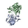
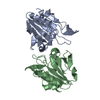
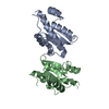
| Title | Escherichia coli Thiol peroxidase (Tpx) wild type disulfide form | ||||||
 Components Components | Thiol peroxidase | ||||||
 Keywords Keywords | OXIDOREDUCTASE / TPX / PEROXIREDOXIN / PEROXIDASE / ANTIOXIDANT | ||||||
| Function / homology |  Function and homology information Function and homology information: / thioredoxin-dependent peroxiredoxin / thioredoxin peroxidase activity / cellular response to oxidative stress / periplasmic space / cytosol Similarity search - Function | ||||||
| Biological species |  | ||||||
| Method |  X-RAY DIFFRACTION / Difference fourier based on isomorphous starting model / Resolution: 2.8 Å X-RAY DIFFRACTION / Difference fourier based on isomorphous starting model / Resolution: 2.8 Å | ||||||
 Authors Authors | Hall, A. / Karplus, P.A. | ||||||
 Citation Citation | Journal: J.Mol.Biol. / Year: 2009 Title: Structural changes common to catalysis in the Tpx peroxiredoxin subfamily. Authors: Hall, A. / Sankaran, B. / Poole, L.B. / Karplus, P.A. | ||||||
| History |
|
- Structure visualization
| Structure viewer | Molecule: MolmilJmol/JSmol |
|---|
- Downloads & links
-Download
| PDBx/mmCIF format | 3i43.cif.gz | 89.1 KB | Display | PDBx/mmCIF format |
|---|---|---|---|---|
| PDB format | pdb3i43.ent.gz | 67.2 KB | Display | PDB format |
| PDBx/mmJSON format | 3i43.json.gz | Tree view | PDBx/mmJSON format | |
| Others |  Other downloads Other downloads |
-Validation report
| Arichive directory | https://data.pdbj.org/pub/pdb/validation_reports/i4/3i43ftp://data.pdbj.org/pub/pdb/validation_reports/i4/3i43 | HTTPS FTP |
|---|
-Related structure data
| Related structure data |  3hvsSC 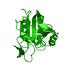 3hvvC  3hvxC S: Starting model for refinement C: citing same article ( |
|---|---|
| Similar structure data |


-Links
 PDBj
PDBj- Assembly
| Deposited unit | 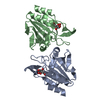
| ||||||||
|---|---|---|---|---|---|---|---|---|---|
| 1 |
| ||||||||
| Unit cell |
|
-Components
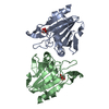
| #1: Protein | Mass: 17717.020 Da / Num. of mol.: 2 Source method: isolated from a genetically manipulated source Details: See Baker and Poole, 2003. / Source: (gene. exp.) #2: Chemical |   Mass: 192.124 Da / Num. of mol.: 2 / Source method: obtained synthetically / Formula: C6H8O7 Mass: 192.124 Da / Num. of mol.: 2 / Source method: obtained synthetically / Formula: C6H8O7#3: Water | ChemComp-HOH / |  Mass: 18.015 Da / Num. of mol.: 602 / Source method: isolated from a natural source / Formula: H2O Mass: 18.015 Da / Num. of mol.: 602 / Source method: isolated from a natural source / Formula: H2OHas protein modification | Y | |
|---|
-Experimental details
-Experiment
| Experiment | Method: X-RAY DIFFRACTION / Number of used crystals: 1 |
|---|
- Sample preparation
| Crystal | Density Matthews: 2.42 Å3/Da / Density % sol: 49.15 % |
|---|---|
| Crystal grow | Temperature: 277 K / Method: vapor diffusion, hanging drop / pH: 4.2 Details: 20% (w/v) PEG-8000, 0.1 M phosphate citrate, 0.2 M sodium chloride, pH 4.2, VAPOR DIFFUSION, HANGING DROP, temperature 277K |
-Data collection
| Diffraction | Mean temperature: 100 K |
|---|---|
| Diffraction source | Source: ROTATING ANODE / Type: RIGAKU RU300 / Wavelength: 1.542 Å |
| Detector | Type: RIGAKU RAXIS IV / Detector: IMAGE PLATE / Date: Nov 28, 2008 / Details: mirrors |
| Radiation | Monochromator: mirrors / Protocol: SINGLE WAVELENGTH / Monochromatic (M) / Laue (L): M / Scattering type: x-ray |
| Radiation wavelength | Wavelength: 1.542 Å / Relative weight: 1 |
| Reflection | Resolution: 2.8→37.36 Å / Num. obs: 7832 / % possible obs: 96.88 % / Redundancy: 2.9 % / Rsym value: 0.105 / Net I/σ(I): 7 |
| Reflection shell | Resolution: 2.8→2.95 Å / Redundancy: 2.9 % / Mean I/σ(I) obs: 2.1 / Num. unique all: 1267 / Rsym value: 0.368 / % possible all: 98 |
- Processing
| Software |
| ||||||||||||||||||||
|---|---|---|---|---|---|---|---|---|---|---|---|---|---|---|---|---|---|---|---|---|---|
| Refinement | Method to determine structure: Difference fourier based on isomorphous starting model Starting model: PDB ENTRY 3HVS Resolution: 2.8→37.36 Å / Cor.coef. Fo:Fc: 0.926 / Cor.coef. Fo:Fc free: 0.91 / SU B: 0.005 / SU ML: 0 / Cross valid method: THROUGHOUT / ESU R: 0.366 / ESU R Free: 0.406 / Stereochemistry target values: MAXIMUM LIKELIHOOD Details: This is not a stand alone structure, but is derived from the higher resolution structure 3HVS by a simple rigid body refinement combined with manual adjustment and geometry optimization of ...Details: This is not a stand alone structure, but is derived from the higher resolution structure 3HVS by a simple rigid body refinement combined with manual adjustment and geometry optimization of only the catalytic residues Cys61 and Cys95. This data set is at much lower resolution and was only used to confirm the presence of an intact disulfide. The conformation of Cys61 and Cys95 from this structure has been added as an alternate conformation with zero occupancy to the higher resolution structure 3HVS and we recommend use of that structure rather than this one.
| ||||||||||||||||||||
| Solvent computation | Ion probe radii: 0.8 Å / Shrinkage radii: 0.8 Å / VDW probe radii: 1.4 Å / Solvent model: MASK | ||||||||||||||||||||
| Displacement parameters | Biso mean: 22.84 Å2
| ||||||||||||||||||||
| Refinement step | Cycle: LAST / Resolution: 2.8→37.36 Å
| ||||||||||||||||||||
| LS refinement shell | Resolution: 2.8→2.872 Å / Total num. of bins used: 20
|
