 Movie
Movie Controller


+ Open data

- Basic information
| Entry | Database: PDB / ID: 3i19 | |||||||||
|---|---|---|---|---|---|---|---|---|---|---|
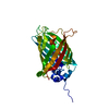
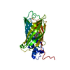
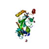
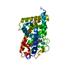
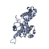
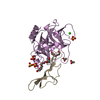
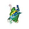
| Title | 1.4 Angstrom Crystal Structure of Fluorescent Protein Cypet | |||||||||
 Components Components | Green fluorescent protein | |||||||||
 Keywords Keywords | FLUORESCENT PROTEIN / BETA BARREL / CHROMOPHORE / LUMINESCENCE / PHOTOPROTEIN | |||||||||
| Function / homology |  Function and homology information Function and homology information | |||||||||
| Biological species |   Aequorea victoria (jellyfish) Aequorea victoria (jellyfish) | |||||||||
| Method |  X-RAY DIFFRACTION / SYNCHROTRON / MOLECULAR REPLACEMENT / Resolution: 1.36 Å X-RAY DIFFRACTION / SYNCHROTRON / MOLECULAR REPLACEMENT / Resolution: 1.36 Å | |||||||||
 Authors Authors | Hu, X. | |||||||||
 Citation Citation | Journal: To be Published Title: 1.4 Angstrom Crystal Structure of Fluorescent Protein Cypet Authors: Liu, R. / Ding, Y. / Hu, X. | |||||||||
| History |
|
- Structure visualization
| Structure viewer | Molecule: MolmilJmol/JSmol |
|---|
- Downloads & links
-Download
| PDBx/mmCIF format | 3i19.cif.gz | 67.5 KB | Display | PDBx/mmCIF format |
|---|---|---|---|---|
| PDB format | pdb3i19.ent.gz | 48.7 KB | Display | PDB format |
| PDBx/mmJSON format | 3i19.json.gz | Tree view | PDBx/mmJSON format | |
| Others |  Other downloads Other downloads |
-Validation report
| Arichive directory | https://data.pdbj.org/pub/pdb/validation_reports/i1/3i19ftp://data.pdbj.org/pub/pdb/validation_reports/i1/3i19 | HTTPS FTP |
|---|
-Related structure data
| Related structure data | 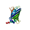 1qyoS S: Starting model for refinement |
|---|---|
| Similar structure data |


-Links
 PDBj
PDBj

- Assembly
| Deposited unit | 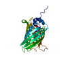
| ||||||||
|---|---|---|---|---|---|---|---|---|---|
| 1 |
| ||||||||
| Unit cell |
| ||||||||
| Details | AUTHOR STATES THAT THE BIOLOGICAL UNIT IS UNKNOWN. |
-Components
| #1: Protein | Mass: 27496.031 Da / Num. of mol.: 1 / Mutation: T9G, V11I, D19E, A87V, I167A, E172T, L194I Source method: isolated from a genetically manipulated source Source: (gene. exp.) Aequorea victoria (jellyfish) / Gene: GFP / Plasmid: pT7His-CyPet / Production host:  |
|---|---|
| #2: Water | ChemComp-HOH /  Mass: 18.015 Da / Num. of mol.: 218 / Source method: isolated from a natural source / Formula: H2O Mass: 18.015 Da / Num. of mol.: 218 / Source method: isolated from a natural source / Formula: H2O |
| Has protein modification | Y |
| Sequence details | THE SEQUENCE WAS MUTATED FROM SER & TYR TO THR & TRP, RESPECTIVELY, TO FORM CHROMOPHORE. THESE FIVE ...THE SEQUENCE WAS MUTATED FROM SER & TYR TO THR & TRP, RESPECTIVE |
-Experimental details
-Experiment
| Experiment | Method: X-RAY DIFFRACTION / Number of used crystals: 1 |
|---|
- Sample preparation
| Crystal | Density Matthews: 2.11 Å3/Da / Density % sol: 41.74 % |
|---|---|
| Crystal grow | Temperature: 293 K / Method: vapor diffusion, hanging drop / pH: 8.5 Details: 30% PEG 4000, 0.1M Tris-HCl pH 8.5, 0.2M Li2SO4, VAPOR DIFFUSION, HANGING DROP, temperature 293K |
-Data collection
| Diffraction | Mean temperature: 100 K |
|---|---|
| Diffraction source | Source: SYNCHROTRON / Site: SSRF  / Beamline: BL17U / Wavelength: 1.0082 Å / Beamline: BL17U / Wavelength: 1.0082 Å |
| Detector | Type: MARMOSAIC 225 mm CCD / Detector: CCD / Date: Jun 16, 2009 |
| Radiation | Protocol: SINGLE WAVELENGTH / Monochromatic (M) / Laue (L): M / Scattering type: x-ray |
| Radiation wavelength | Wavelength: 1.0082 Å / Relative weight: 1 |
| Reflection | Resolution: 1.36→50 Å / Num. obs: 49295 / % possible obs: 99 % / Observed criterion σ(F): 2 / Observed criterion σ(I): 2 / Redundancy: 5.8 % / Rmerge(I) obs: 0.047 / Rsym value: 0.042 / Net I/σ(I): 41 / Num. measured all: 550430 |
| Reflection shell | Resolution: 1.36→1.38 Å / Redundancy: 4.5 % / Rmerge(I) obs: 0.194 / Mean I/σ(I) obs: 6.1 / Num. unique all: 2238 / Rsym value: 0.201 / % possible all: 91.6 |
- Processing
| Software |
| ||||||||||||||||||||||||||||||||||||||||||||||||||||||||||||||||||||||
|---|---|---|---|---|---|---|---|---|---|---|---|---|---|---|---|---|---|---|---|---|---|---|---|---|---|---|---|---|---|---|---|---|---|---|---|---|---|---|---|---|---|---|---|---|---|---|---|---|---|---|---|---|---|---|---|---|---|---|---|---|---|---|---|---|---|---|---|---|---|---|---|
| Refinement | Method to determine structure: MOLECULAR REPLACEMENT Starting model: PDB ENTRY 1QYO Resolution: 1.36→29.14 Å / Cor.coef. Fo:Fc: 0.962 / Cor.coef. Fo:Fc free: 0.957 / SU B: 0.726 / SU ML: 0.031 / Isotropic thermal model: overall / Cross valid method: THROUGHOUT / σ(F): 2 / σ(I): 2 / ESU R: 0.056 / ESU R Free: 0.06 / Stereochemistry target values: MAXIMUM LIKELIHOOD
| ||||||||||||||||||||||||||||||||||||||||||||||||||||||||||||||||||||||
| Solvent computation | Ion probe radii: 0.8 Å / Shrinkage radii: 0.8 Å / VDW probe radii: 1.2 Å / Solvent model: MASK | ||||||||||||||||||||||||||||||||||||||||||||||||||||||||||||||||||||||
| Displacement parameters | Biso mean: 15.314 Å2
| ||||||||||||||||||||||||||||||||||||||||||||||||||||||||||||||||||||||
| Refinement step | Cycle: LAST / Resolution: 1.36→29.14 Å
| ||||||||||||||||||||||||||||||||||||||||||||||||||||||||||||||||||||||
| Refine LS restraints |
| ||||||||||||||||||||||||||||||||||||||||||||||||||||||||||||||||||||||
| LS refinement shell | Resolution: 1.36→1.396 Å / Total num. of bins used: 20
|
