- PDB-3i10: Crystal structure of Putative glycerophosphoryl diester phosphodi... -
+
Open data
ID or keywords:
Loading...
-
Basic information
Entry
Database: PDB / ID: 3i10
Title
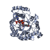
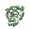
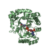
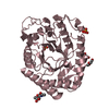
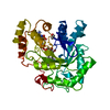
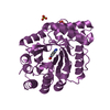
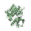
Crystal structure of Putative glycerophosphoryl diester phosphodiesterase (NP_812074.1) from BACTEROIDES THETAIOTAOMICRON VPI-5482 at 1.35 A resolution
Mass: 18.015 Da / Num. of mol.: 356 / Source method: isolated from a natural source / Formula: H2O
Has protein modification
Y
Sequence details
SEQUENCE THIS CONSTRUCT (RESIDUES 25-301) WAS EXPRESSED WITH A PURIFICATION TAG MGSDKIHHHHHHENLYFQG. ...SEQUENCE THIS CONSTRUCT (RESIDUES 25-301) WAS EXPRESSED WITH A PURIFICATION TAG MGSDKIHHHHHHENLYFQG. THE TAG WAS REMOVED WITH TEV PROTEASE LEAVING ONLY A GLYCINE (0) FOLLOWED BY THE TARGET SEQUENCE.
-
Experimental details
-
Experiment
Experiment
Method: X-RAY DIFFRACTION / Number of used crystals: 1
-
Sample preparation
Crystal
Density Matthews: 2.18 Å3/Da / Density % sol: 43.53 %
Crystal grow
Temperature: 277 K / Method: vapor diffusion, sitting drop / pH: 6.5 Details: 0.2000M NaOAc, 30.0000% PEG-8000, 0.1M Cacodylate pH 6.5, NANODROP, VAPOR DIFFUSION, SITTING DROP, temperature 277K
Type: MARMOSAIC 325 mm CCD / Detector: CCD / Date: Mar 27, 2009 / Details: Flat mirror (vertical focusing)
Radiation
Monochromator: Single crystal Si(111) bent monochromator (horizontal focusing) Protocol: MAD / Monochromatic (M) / Laue (L): M / Scattering type: x-ray
Radiation wavelength
ID
Wavelength (Å)
Relative weight
1
0.91837
1
2
0.97993
1
3
0.9792
1
Reflection
Resolution: 1.35→29.05 Å / Num. obs: 61313 / % possible obs: 100 % / Redundancy: 3.6 % / Biso Wilson estimate: 10.639 Å2 / Rmerge(I) obs: 0.085 / Rsym value: 0.085 / Net I/σ(I): 9.7
Reflection shell
Diffraction-ID: 1
Resolution (Å)
Redundancy (%)
Rmerge(I) obs
Mean I/σ(I) obs
Num. measured all
Num. unique all
Rsym value
% possible all
1.35-1.39
3.1
0.59
1.9
14135
4503
0.59
100
1.39-1.42
3.6
0.525
2.3
15862
4379
0.525
100
1.42-1.46
3.6
0.438
2.8
15316
4212
0.438
100
1.46-1.51
3.6
0.359
3.4
15137
4151
0.359
100
1.51-1.56
3.7
0.29
4.3
14626
4001
0.29
100
1.56-1.61
3.7
0.253
4.9
14185
3875
0.253
100
1.61-1.67
3.7
0.212
5.8
13782
3762
0.212
100
1.67-1.74
3.7
0.183
6.9
13396
3641
0.183
100
1.74-1.82
3.7
0.146
8.6
12669
3449
0.146
100
1.82-1.91
3.7
0.114
10.5
12182
3301
0.114
100
1.91-2.01
3.7
0.092
12.5
11765
3182
0.092
100
2.01-2.13
3.7
0.078
14.2
11143
3030
0.078
100
2.13-2.28
3.7
0.078
16
10358
2806
0.078
100
2.28-2.46
3.7
0.079
18
9756
2649
0.079
100
2.46-2.7
3.7
0.067
19.2
8908
2415
0.067
100
2.7-3.02
3.7
0.056
20.5
8191
2227
0.056
100
3.02-3.49
3.7
0.046
22.9
7151
1958
0.046
100
3.49-4.27
3.6
0.043
25
6183
1700
0.043
100
4.27-6.04
3.6
0.043
24.3
4694
1317
0.043
100
6.04-29.05
3.3
0.045
23.2
2525
755
0.045
98.6
-
Phasing
Phasing
Method: MAD
-
Processing
Software
Name
Version
Classification
NB
REFMAC
5.5.0053
refinement
PHENIX
refinement
SHELX
phasing
MolProbity
3beta29
modelbuilding
SCALA
3.2.5
datascaling
PDB_EXTRACT
3.006
dataextraction
MOSFLM
datareduction
SHELXD
phasing
autoSHARP
phasing
Refinement
Method to determine structure: MAD / Resolution: 1.35→29.05 Å / Cor.coef. Fo:Fc: 0.972 / Cor.coef. Fo:Fc free: 0.962 / Occupancy max: 1 / Occupancy min: 0.15 / SU B: 1.738 / SU ML: 0.032 / TLS residual ADP flag: LIKELY RESIDUAL / Cross valid method: THROUGHOUT / σ(F): 0 / ESU R: 0.051 / ESU R Free: 0.053 Stereochemistry target values: MAXIMUM LIKELIHOOD WITH PHASES Details: 1. HYDROGENS HAVE BEEN ADDED IN THE RIDING POSITIONS. 2. ATOM RECORD CONTAINS RESIDUAL B FACTORS ONLY. 3. A MET-INHIBITION PROTOCOL WAS USED FOR SELENOMETHIONINE INCORPORATION DURING PROTEIN ...Details: 1. HYDROGENS HAVE BEEN ADDED IN THE RIDING POSITIONS. 2. ATOM RECORD CONTAINS RESIDUAL B FACTORS ONLY. 3. A MET-INHIBITION PROTOCOL WAS USED FOR SELENOMETHIONINE INCORPORATION DURING PROTEIN EXPRESSION. THE OCCUPANCY OF THE SE ATOMS IN THE MSE RESIDUES WAS REDUCED TO 0.75 TO ACCOUNT FOR THE REDUCED SCATTERING POWER DUE TO PARTIAL S-MET INCORPORATION. 4. ACETATE (ACT), ETHYLENE GLYCOL (EDO), AND POLYETHYLENE GLYCOL (PEG) FROM THE CRYSTALLIZATION/CRYOPROTECTANT SOLUTIONS HAVE BEEN MODELED INTO THE SOLVENT STRUCTURE. 5. TLS GROUPS WERE ASSIGNED WITH THE AID OF THE TLSMD SERVER.
Rfactor
Num. reflection
% reflection
Selection details
Rfree
0.172
3109
5.1 %
RANDOM
Rwork
0.15
-
-
-
obs
0.151
61269
99.96 %
-
Solvent computation
Ion probe radii: 0.8 Å / Shrinkage radii: 0.8 Å / VDW probe radii: 1.4 Å / Solvent model: MASK
In the structure databanks used in Yorodumi, some data are registered as the other names, "COVID-19 virus" and "2019-nCoV". Here are the details of the virus and the list of structure data.
Jan 31, 2019. EMDB accession codes are about to change! (news from PDBe EMDB page)
EMDB accession codes are about to change! (news from PDBe EMDB page)
The allocation of 4 digits for EMDB accession codes will soon come to an end. Whilst these codes will remain in use, new EMDB accession codes will include an additional digit and will expand incrementally as the available range of codes is exhausted. The current 4-digit format prefixed with “EMD-” (i.e. EMD-XXXX) will advance to a 5-digit format (i.e. EMD-XXXXX), and so on. It is currently estimated that the 4-digit codes will be depleted around Spring 2019, at which point the 5-digit format will come into force.
The EM Navigator/Yorodumi systems omit the EMD- prefix.
Related info.:Q: What is EMD? / ID/Accession-code notation in Yorodumi/EM Navigator
Yorodumi is a browser for structure data from EMDB, PDB, SASBDB, etc.
This page is also the successor to EM Navigator detail page, and also detail information page/front-end page for Omokage search.
The word "yorodu" (or yorozu) is an old Japanese word meaning "ten thousand". "mi" (miru) is to see.
Related info.:EMDB / PDB / SASBDB / Comparison of 3 databanks / Yorodumi Search / Aug 31, 2016. New EM Navigator & Yorodumi / Yorodumi Papers / Jmol/JSmol / Function and homology information / Changes in new EM Navigator and Yorodumi
 Movie
Movie Controller

 Yorodumi
 Open data
 Basic information
 Components
 Keywords
 Function and homology information
 Bacteroides thetaiotaomicron VPI-5482 (bacteria)
 X-RAY DIFFRACTION /
  Authors
 Citation
 Structure visualization
 Downloads & links
 Other downloads
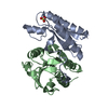


 PDBj
PDBj Assembly
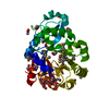

 Mass: 59.044 Da / Num. of mol.: 2 / Source method: obtained synthetically / Formula: C2H3O2
 Mass: 62.068 Da / Num. of mol.: 18 / Source method: obtained synthetically / Formula: C2H6O2
 Mass: 106.120 Da / Num. of mol.: 1 / Source method: obtained synthetically / Formula: C4H10O3
 Mass: 18.015 Da / Num. of mol.: 356 / Source method: isolated from a natural source / Formula: H2O
 Sample preparation
 / Beamline: BL11-1 / Wavelength: 0.91837,0.97993,0.97920
 Processing