 Movie
Movie Controller

[English] 日本語
 Yorodumi
- PDB-3hzz: 2.4 Angstrom Crystal Structure of Streptomyces collinus crotonyl ... -
+ Open data

- Basic information
| Entry | Database: PDB / ID: 3hzz | ||||||
|---|---|---|---|---|---|---|---|
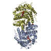
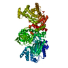
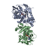
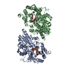
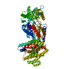
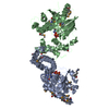
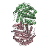
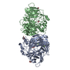
| Title | 2.4 Angstrom Crystal Structure of Streptomyces collinus crotonyl CoA carboxylase/reductase | ||||||
 Components Components | Crotonyl CoA reductase | ||||||
 Keywords Keywords | OXIDOREDUCTASE / Reductase / Carboxylase / alcohol dehydrogenase / biocatalysis / enoyl reductase / glyoxolate cycle / acetyl CoA assimilation / methylotrophy / serine cycle / polyketide | ||||||
| Function / homology |  Function and homology information Function and homology informationcrotonyl-CoA reductase / crotonyl-CoA reductase activity / NADP binding / metal ion binding Similarity search - Function | ||||||
| Biological species |  Streptomyces collinus (bacteria) Streptomyces collinus (bacteria) | ||||||
| Method |  X-RAY DIFFRACTION / MIR / Resolution: 2.4 Å X-RAY DIFFRACTION / MIR / Resolution: 2.4 Å | ||||||
 Authors Authors | Scarsdale, J.N. / Musayev, F.N. / Wright, H.T. | ||||||
 Citation Citation | Journal: To be Published Title: Structure of Streptomycs collinus crotonyl COA carboxylase/reductase Authors: Scarsdale, J.N. / Musayev, F.N. / Hazzard, C. / Florova, G. / Reynolds, K. / Wright, H.T. #1: Journal: Proc.Natl.Acad.Sci.USA / Year: 2009 Title: Carboxylation mechanism and stereochemistry of crotonyl-CoA carboxylase/reductase, a carboxylating enoyl-thioester reductase Authors: Erb, T.J. / Brecht, V. / Fuchs, G. / Muller, M. / Alber, B.E. #2: Journal: Proc.Natl.Acad.Sci.USA / Year: 2007 Title: Synthesis of C5-dicarboxylic acids from C2-units involving crotonyl-CoA carboxylase/reductase: The ethylmalonyl-CoA pathway Authors: Erb, T.J. / Berg, I.A. / Brecht, V. / Muller, M. / Fuchs, G. / Alber, B.E. | ||||||
| History |
|
- Structure visualization
| Structure viewer | Molecule: MolmilJmol/JSmol |
|---|
- Downloads & links
-Download
| PDBx/mmCIF format | 3hzz.cif.gz | 676.8 KB | Display | PDBx/mmCIF format |
|---|---|---|---|---|
| PDB format | pdb3hzz.ent.gz | 562.6 KB | Display | PDB format |
| PDBx/mmJSON format | 3hzz.json.gz | Tree view | PDBx/mmJSON format | |
| Others |  Other downloads Other downloads |
-Validation report
| Arichive directory | https://data.pdbj.org/pub/pdb/validation_reports/hz/3hzzftp://data.pdbj.org/pub/pdb/validation_reports/hz/3hzz | HTTPS FTP |
|---|
-Related structure data
| Similar structure data |
|---|


-Links
 PDBj
PDBj

- Assembly
| Deposited unit | 
| ||||||||
|---|---|---|---|---|---|---|---|---|---|
| 1 | 
| ||||||||
| 2 | 
| ||||||||
| 3 | 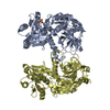
| ||||||||
| 4 | 
| ||||||||
| 5 |
| ||||||||
| Unit cell |
| ||||||||
| Details | Author-defined biological assembly is based on the fact that the dimer interface for dimers (A+B) as well as (C+D) is in a beta sheet. Size-exclusion chromatography indicates protein runs as a dimer |
-Components
| #1: Protein | Mass: 51597.969 Da / Num. of mol.: 4 Source method: isolated from a genetically manipulated source Source: (gene. exp.) Streptomyces collinus (bacteria) / Gene: ccr, crotonyl COA reductase / Plasmid: pET28a (Novagen) / Production host: #2: Chemical | ChemComp-SO4 /   Mass: 96.063 Da / Num. of mol.: 5 / Source method: obtained synthetically / Formula: SO4 Mass: 96.063 Da / Num. of mol.: 5 / Source method: obtained synthetically / Formula: SO4#3: Water | ChemComp-HOH / |  Mass: 18.015 Da / Num. of mol.: 585 / Source method: isolated from a natural source / Formula: H2O Mass: 18.015 Da / Num. of mol.: 585 / Source method: isolated from a natural source / Formula: H2O |
|---|
-Experimental details
-Experiment
| Experiment | Method: X-RAY DIFFRACTION / Number of used crystals: 1 |
|---|
- Sample preparation
| Crystal | Density Matthews: 2.53 Å3/Da / Density % sol: 51.39 % |
|---|---|
| Crystal grow | Temperature: 293 K / Method: vapor diffusion with microseeding / pH: 6.2 Details: 0.1M MES, 2.0M Ammonium Sulfate, 3% PEG 200, pH 6.2, vapor diffusion with microseeding, temperature 293K |
-Data collection
| Diffraction | Mean temperature: 100 K |
|---|---|
| Diffraction source | Source: ROTATING ANODE / Type: RIGAKU MICROMAX-007 / Wavelength: 1.5418 Å |
| Detector | Type: RIGAKU RAXIS IV++ / Detector: IMAGE PLATE / Date: Jul 13, 2008 / Details: Rigaku Varimax confocal optics |
| Radiation | Monochromator: Rigaku Varimax Confocal optics / Protocol: SINGLE WAVELENGTH / Monochromatic (M) / Laue (L): M / Scattering type: x-ray |
| Radiation wavelength | Wavelength: 1.5418 Å / Relative weight: 1 |
| Reflection | Resolution: 2.4→17.97 Å / Num. all: 82617 / Num. obs: 78199 / % possible obs: 94.7 % / Redundancy: 11.26 % / Biso Wilson estimate: 38.5 Å2 / Rmerge(I) obs: 0.119 / Net I/σ(I): 15.3 |
| Reflection shell | Resolution: 2.4→2.49 Å / Redundancy: 11.08 % / Rmerge(I) obs: 0.344 / Mean I/σ(I) obs: 7.4 / Num. unique all: 8154 / Rsym value: 0.344 / % possible all: 92.5 |
- Processing
| Software |
| ||||||||||||||||||||||||||||||||||||||||||||||||||||||||||||||||||||||||||||||||||||||||||||||||||||||||||||||||||||||||||||||||||||||||||||||||||||||||||||||||||||||||||
|---|---|---|---|---|---|---|---|---|---|---|---|---|---|---|---|---|---|---|---|---|---|---|---|---|---|---|---|---|---|---|---|---|---|---|---|---|---|---|---|---|---|---|---|---|---|---|---|---|---|---|---|---|---|---|---|---|---|---|---|---|---|---|---|---|---|---|---|---|---|---|---|---|---|---|---|---|---|---|---|---|---|---|---|---|---|---|---|---|---|---|---|---|---|---|---|---|---|---|---|---|---|---|---|---|---|---|---|---|---|---|---|---|---|---|---|---|---|---|---|---|---|---|---|---|---|---|---|---|---|---|---|---|---|---|---|---|---|---|---|---|---|---|---|---|---|---|---|---|---|---|---|---|---|---|---|---|---|---|---|---|---|---|---|---|---|---|---|---|---|---|---|
| Refinement | Method to determine structure: MIR / Resolution: 2.4→17.97 Å / Cor.coef. Fo:Fc: 0.93 / Cor.coef. Fo:Fc free: 0.882 / SU B: 21.702 / SU ML: 0.219 Isotropic thermal model: TLS with individual residual isotropic B factors Cross valid method: THROUGHOUT / ESU R: 0.446 / ESU R Free: 0.275 / Stereochemistry target values: MAXIMUM LIKELIHOOD
| ||||||||||||||||||||||||||||||||||||||||||||||||||||||||||||||||||||||||||||||||||||||||||||||||||||||||||||||||||||||||||||||||||||||||||||||||||||||||||||||||||||||||||
| Solvent computation | Ion probe radii: 0.8 Å / Shrinkage radii: 0.8 Å / VDW probe radii: 1.4 Å / Solvent model: BABINET MODEL WITH MASK | ||||||||||||||||||||||||||||||||||||||||||||||||||||||||||||||||||||||||||||||||||||||||||||||||||||||||||||||||||||||||||||||||||||||||||||||||||||||||||||||||||||||||||
| Displacement parameters | Biso mean: 34.779 Å2
| ||||||||||||||||||||||||||||||||||||||||||||||||||||||||||||||||||||||||||||||||||||||||||||||||||||||||||||||||||||||||||||||||||||||||||||||||||||||||||||||||||||||||||
| Refine analyze | Luzzati coordinate error obs: 0.35 Å | ||||||||||||||||||||||||||||||||||||||||||||||||||||||||||||||||||||||||||||||||||||||||||||||||||||||||||||||||||||||||||||||||||||||||||||||||||||||||||||||||||||||||||
| Refinement step | Cycle: LAST / Resolution: 2.4→17.97 Å
| ||||||||||||||||||||||||||||||||||||||||||||||||||||||||||||||||||||||||||||||||||||||||||||||||||||||||||||||||||||||||||||||||||||||||||||||||||||||||||||||||||||||||||
| Refine LS restraints |
| ||||||||||||||||||||||||||||||||||||||||||||||||||||||||||||||||||||||||||||||||||||||||||||||||||||||||||||||||||||||||||||||||||||||||||||||||||||||||||||||||||||||||||
| LS refinement shell | Resolution: 2.4→2.462 Å / Total num. of bins used: 20
| ||||||||||||||||||||||||||||||||||||||||||||||||||||||||||||||||||||||||||||||||||||||||||||||||||||||||||||||||||||||||||||||||||||||||||||||||||||||||||||||||||||||||||
| Refinement TLS params. | Method: refined / Refine-ID: X-RAY DIFFRACTION
| ||||||||||||||||||||||||||||||||||||||||||||||||||||||||||||||||||||||||||||||||||||||||||||||||||||||||||||||||||||||||||||||||||||||||||||||||||||||||||||||||||||||||||
| Refinement TLS group |
|
