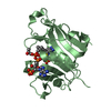
Resolution: 1.8→46.3 Å / Cor.coef. Fo:Fc: 0.944 / Cor.coef. Fo:Fc free: 0.938 / SU B: 0.003 / SU ML: 0 / Cross valid method: THROUGHOUT / ESU R: 0.118 / ESU R Free: 0.127 / Stereochemistry target values: MAXIMUM LIKELIHOOD Details: HYDROGENS HAVE BEEN ADDED IN THE RIDING POSITIONS: THE LIGAND 10F IN THIS ENTRY IS 10-FORMYLTETRAHYDROFOLATE. HOWEVER, THERE IS SOME DISCREPANCY IN THE GEOMETRY. THE GEOMETRY FOR 10F IS ...Details: HYDROGENS HAVE BEEN ADDED IN THE RIDING POSITIONS: THE LIGAND 10F IN THIS ENTRY IS 10-FORMYLTETRAHYDROFOLATE. HOWEVER, THERE IS SOME DISCREPANCY IN THE GEOMETRY. THE GEOMETRY FOR 10F IS SUGGESTED BY THE REFINEMENT. THE CO-ORDINATES FIT WELL IN THE ELECTRON DENSITY MAP. THE MAP WAS GENERATED USING A DATASET COLLECTED AT 1.8 ANGSTROM RESOLUTION. THE DENSITY FOR THE LIGAND IS UNAMBIGUOUS AND THEREFORE THE GEOMETRIES ARE CORRECT AND ARE AS THEY WOULD BE IN A BIOLOGICAL MOLECULE, WHERE THE MICRO ENVIRONMENT HAS A PROFOUND INFLUENCE ON THE GEOMETRIES OF THE LIGAND.
Rfactor
Num. reflection
% reflection
Selection details
Rfree
0.23184
1027
5.1 %
RANDOM
Rwork
0.21538
-
-
-
all
0.21624
23583
-
-
obs
0.21624
18978
99.94 %
-
Solvent computation
Ion probe radii: 0.8 Å / Shrinkage radii: 0.8 Å / VDW probe radii: 1.4 Å / Solvent model: MASK
Displacement parameters
Biso mean: 26.119 Å2
Baniso -1
Baniso -2
Baniso -3
1-
1.59 Å2
0 Å2
0 Å2
2-
-
-0.32 Å2
0 Å2
3-
-
-
-1.27 Å2
Refinement step
Cycle: LAST / Resolution: 1.8→46.3 Å
Protein
Nucleic acid
Ligand
Solvent
Total
Num. atoms
1521
0
45
137
1703
Refine LS restraints
Refine-ID
Type
Dev ideal
X-RAY DIFFRACTION
r_bond_d
0.028
X-RAY DIFFRACTION
r_angle_deg
2.119
LS refinement shell
Resolution: 1.8→1.847 Å / Total num. of bins used: 20
Rfactor
Num. reflection
% reflection
Rfree
0.306
78
-
Rwork
0.258
1362
-
obs
-
-
100 %
+
About Yorodumi
-
News
-
Feb 9, 2022. New format data for meta-information of EMDB entries
New format data for meta-information of EMDB entries
Version 3 of the EMDB header file is now the official format.
The previous official version 1.9 will be removed from the archive.
In the structure databanks used in Yorodumi, some data are registered as the other names, "COVID-19 virus" and "2019-nCoV". Here are the details of the virus and the list of structure data.
Jan 31, 2019. EMDB accession codes are about to change! (news from PDBe EMDB page)
EMDB accession codes are about to change! (news from PDBe EMDB page)
The allocation of 4 digits for EMDB accession codes will soon come to an end. Whilst these codes will remain in use, new EMDB accession codes will include an additional digit and will expand incrementally as the available range of codes is exhausted. The current 4-digit format prefixed with “EMD-” (i.e. EMD-XXXX) will advance to a 5-digit format (i.e. EMD-XXXXX), and so on. It is currently estimated that the 4-digit codes will be depleted around Spring 2019, at which point the 5-digit format will come into force.
The EM Navigator/Yorodumi systems omit the EMD- prefix.
Related info.:Q: What is EMD? / ID/Accession-code notation in Yorodumi/EM Navigator
Yorodumi is a browser for structure data from EMDB, PDB, SASBDB, etc.
This page is also the successor to EM Navigator detail page, and also detail information page/front-end page for Omokage search.
The word "yorodu" (or yorozu) is an old Japanese word meaning "ten thousand". "mi" (miru) is to see.
Related info.:EMDB / PDB / SASBDB / Comparison of 3 databanks / Yorodumi Search / Aug 31, 2016. New EM Navigator & Yorodumi / Yorodumi Papers / Jmol/JSmol / Function and homology information / Changes in new EM Navigator and Yorodumi
 Movie
Movie Controller


 Open data
 Basic information
 Components
 Keywords
 Function and homology information
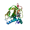
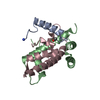
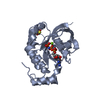
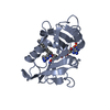
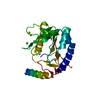
 Homo sapiens (human)
 X-RAY DIFFRACTION /
  Authors
 Citation
 Structure visualization
 Downloads & links
 Other downloads


 PDBj
PDBj

 Assembly
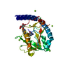


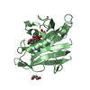
 Mass: 479.487 Da / Num. of mol.: 1 / Source method: obtained synthetically / Formula: C20H29N7O7
 Mass: 58.693 Da / Num. of mol.: 2 / Source method: obtained synthetically / Formula: Ni
 Mass: 24.305 Da / Num. of mol.: 9 / Source method: obtained synthetically / Formula: Mg
 Mass: 18.015 Da / Num. of mol.: 137 / Source method: isolated from a natural source / Formula: H2O
 Sample preparation
 / Beamline: 19-ID / Wavelength: 0.979 Å
 Processing