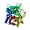
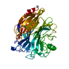
 Movie
Movie Controller


+ Open data

- Basic information
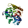
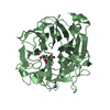
| Entry | Database: PDB / ID: 3hvm | ||||||
|---|---|---|---|---|---|---|---|
| Title | Agmatine Deiminase from Helicobacter pylori | ||||||
 Components Components | AGMATINE DEIMINASE | ||||||
 Keywords Keywords | HYDROLASE / Agmatine Deiminase | ||||||
| Function / homology |  Function and homology information Function and homology informationagmatine deiminase activity / putrescine biosynthetic process / protein-arginine deiminase activity Similarity search - Function | ||||||
| Biological species |   Helicobacter pylori (bacteria) Helicobacter pylori (bacteria) | ||||||
| Method |  X-RAY DIFFRACTION / SYNCHROTRON / MOLECULAR REPLACEMENT / Resolution: 2.1 Å X-RAY DIFFRACTION / SYNCHROTRON / MOLECULAR REPLACEMENT / Resolution: 2.1 Å | ||||||
 Authors Authors | Jones, J. / Lovelace, L. / Lebioda, L. / Thompson, P. | ||||||
 Citation Citation | Journal: Bioorg.Chem. / Year: 2010 Title: Characterization and inactivation of an agmatine deiminase from Helicobacter pylori. Authors: Jones, J.E. / Causey, C.P. / Lovelace, L. / Knuckley, B. / Flick, H. / Lebioda, L. / Thompson, P.R. | ||||||
| History |
|
- Structure visualization
| Structure viewer | Molecule: MolmilJmol/JSmol |
|---|
- Downloads & links
-Download
| PDBx/mmCIF format | 3hvm.cif.gz | 82.3 KB | Display | PDBx/mmCIF format |
|---|---|---|---|---|
| PDB format | pdb3hvm.ent.gz | 60.4 KB | Display | PDB format |
| PDBx/mmJSON format | 3hvm.json.gz | Tree view | PDBx/mmJSON format | |
| Others |  Other downloads Other downloads |
-Validation report
| Arichive directory | https://data.pdbj.org/pub/pdb/validation_reports/hv/3hvmftp://data.pdbj.org/pub/pdb/validation_reports/hv/3hvm | HTTPS FTP |
|---|
-Related structure data
| Related structure data |  2cmuS S: Starting model for refinement |
|---|---|
| Similar structure data |


-Links
 PDBj
PDBj- Assembly
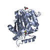
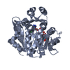
| Deposited unit | 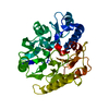
| ||||||||
|---|---|---|---|---|---|---|---|---|---|
| 1 |
| ||||||||
| Unit cell |
|
-Components
| #1: Protein | Mass: 37614.129 Da / Num. of mol.: 1 Source method: isolated from a genetically manipulated source Source: (gene. exp.) Helicobacter pylori (bacteria) / Strain: J99 / Gene: HPAG1_0045, jhp_0042 / Production host: |
|---|---|
| #2: Water | ChemComp-HOH /  Mass: 18.015 Da / Num. of mol.: 164 / Source method: isolated from a natural source / Formula: H2O Mass: 18.015 Da / Num. of mol.: 164 / Source method: isolated from a natural source / Formula: H2O |
| Sequence details | MUTATIONS OCCURRED DURING CLONING OF HPAGD |
-Experimental details
-Experiment
| Experiment | Method: X-RAY DIFFRACTION / Number of used crystals: 1 |
|---|
- Sample preparation
| Crystal | Density Matthews: 2.56 Å3/Da / Density % sol: 52.04 % |
|---|---|
| Crystal grow | Temperature: 298 K / Method: vapor diffusion, hanging drop / pH: 5.6 Details: 15% PEG 4K, 10 mM sodium citrate pH 5.6, and 7% isopropanol, VAPOR DIFFUSION, HANGING DROP, temperature 298K |
-Data collection
| Diffraction | Mean temperature: 100 K |
|---|---|
| Diffraction source | Source: SYNCHROTRON / Site: APS  / Beamline: 22-ID / Wavelength: 1 Å / Beamline: 22-ID / Wavelength: 1 Å |
| Detector | Type: MAR scanner 300 mm plate / Detector: IMAGE PLATE / Date: Feb 24, 2007 |
| Radiation | Monochromator: Si 220 / Protocol: SINGLE WAVELENGTH / Monochromatic (M) / Laue (L): M / Scattering type: x-ray |
| Radiation wavelength | Wavelength: 1 Å / Relative weight: 1 |
| Reflection | Resolution: 2.1→30.37 Å / Num. obs: 18081 / % possible obs: 76 % / Redundancy: 3.6 % / Rmerge(I) obs: 0.043 / Χ2: 1.374 / Net I/σ(I): 31.053 |
| Reflection shell | Resolution: 2.1→2.18 Å / Redundancy: 3.2 % / Rmerge(I) obs: 0.143 / Mean I/σ(I) obs: 16.5 / Num. unique all: 1384 / Χ2: 0.82 / % possible all: 59.8 |
- Processing
| Software |
| ||||||||||||||||||||||||||||||||||||||||||||||||||||||||||||||||||||||||||||||||||||||||||
|---|---|---|---|---|---|---|---|---|---|---|---|---|---|---|---|---|---|---|---|---|---|---|---|---|---|---|---|---|---|---|---|---|---|---|---|---|---|---|---|---|---|---|---|---|---|---|---|---|---|---|---|---|---|---|---|---|---|---|---|---|---|---|---|---|---|---|---|---|---|---|---|---|---|---|---|---|---|---|---|---|---|---|---|---|---|---|---|---|---|---|---|
| Refinement | Method to determine structure: MOLECULAR REPLACEMENT Starting model: 2CMU Resolution: 2.1→30.37 Å / Cor.coef. Fo:Fc: 0.94 / Cor.coef. Fo:Fc free: 0.91 / WRfactor Rfree: 0.233 / WRfactor Rwork: 0.226 / Occupancy max: 1 / Occupancy min: 1 / FOM work R set: 0.84 / SU B: 9.861 / SU ML: 0.121 / SU R Cruickshank DPI: 0.374 / SU Rfree: 0.228 / TLS residual ADP flag: LIKELY RESIDUAL / Cross valid method: THROUGHOUT / σ(F): 0 / ESU R: 0.324 / ESU R Free: 0.246 / Stereochemistry target values: MAXIMUM LIKELIHOOD / Details: HYDROGENS HAVE BEEN ADDED IN THE RIDING POSITIONS
| ||||||||||||||||||||||||||||||||||||||||||||||||||||||||||||||||||||||||||||||||||||||||||
| Solvent computation | Ion probe radii: 0.8 Å / Shrinkage radii: 0.8 Å / VDW probe radii: 1.2 Å / Solvent model: MASK | ||||||||||||||||||||||||||||||||||||||||||||||||||||||||||||||||||||||||||||||||||||||||||
| Displacement parameters | Biso max: 63.93 Å2 / Biso mean: 29.966 Å2 / Biso min: 8.97 Å2
| ||||||||||||||||||||||||||||||||||||||||||||||||||||||||||||||||||||||||||||||||||||||||||
| Refinement step | Cycle: LAST / Resolution: 2.1→30.37 Å
| ||||||||||||||||||||||||||||||||||||||||||||||||||||||||||||||||||||||||||||||||||||||||||
| Refine LS restraints |
| ||||||||||||||||||||||||||||||||||||||||||||||||||||||||||||||||||||||||||||||||||||||||||
| LS refinement shell | Resolution: 2.1→2.156 Å / Total num. of bins used: 20
| ||||||||||||||||||||||||||||||||||||||||||||||||||||||||||||||||||||||||||||||||||||||||||
| Refinement TLS params. | Method: refined / Origin x: -8.5798 Å / Origin y: -0.6163 Å / Origin z: -17.9363 Å
| ||||||||||||||||||||||||||||||||||||||||||||||||||||||||||||||||||||||||||||||||||||||||||
| Refinement TLS group |
|
