 Movie
Movie Controller

[English] 日本語
 Yorodumi
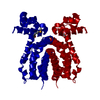
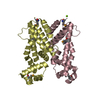
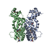
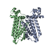
- PDB-3htj: Crystal structure of multidrug binding protein EbrR complexed wit... -
+ Open data

- Basic information
| Entry | Database: PDB / ID: 3htj | ||||||
|---|---|---|---|---|---|---|---|
| Title | Crystal structure of multidrug binding protein EbrR complexed with ethidium | ||||||
 Components Components | EbrA repressor | ||||||
 Keywords Keywords | DNA BINDING PROTEIN / TetR family / multidrug resistance / Multidrug binding protein / DNA-binding / Transcription / Transcription regulation | ||||||
| Function / homology |  Function and homology information Function and homology informationtranscription cis-regulatory region binding / DNA-binding transcription factor activity Similarity search - Function | ||||||
| Biological species |  Streptomyces lividans (bacteria) Streptomyces lividans (bacteria) | ||||||
| Method |  X-RAY DIFFRACTION / SYNCHROTRON / MOLECULAR REPLACEMENT / Resolution: 2.7 Å X-RAY DIFFRACTION / SYNCHROTRON / MOLECULAR REPLACEMENT / Resolution: 2.7 Å | ||||||
 Authors Authors | Dong, J. / Ni, L. / Schumacher, M. / Brennan, R. | ||||||
 Citation Citation | Journal: To be Published Title: Structural plasticity is key to multiple ligand binding by the multidrug binding regulator EbrR Authors: Dong, J. / Ni, L. / Schumacher, M. / Brennan, R. | ||||||
| History |
|
- Structure visualization
| Structure viewer | Molecule: MolmilJmol/JSmol |
|---|
- Downloads & links
-Download
| PDBx/mmCIF format | 3htj.cif.gz | 80.6 KB | Display | PDBx/mmCIF format |
|---|---|---|---|---|
| PDB format | pdb3htj.ent.gz | 59.6 KB | Display | PDB format |
| PDBx/mmJSON format | 3htj.json.gz | Tree view | PDBx/mmJSON format | |
| Others |  Other downloads Other downloads |
-Validation report
| Arichive directory | https://data.pdbj.org/pub/pdb/validation_reports/ht/3htjftp://data.pdbj.org/pub/pdb/validation_reports/ht/3htj | HTTPS FTP |
|---|
-Related structure data
| Related structure data |  3htaSC 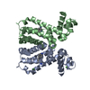 3hthC 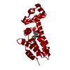 3htiC S: Starting model for refinement C: citing same article ( |
|---|---|
| Similar structure data |


-Links
 PDBj
PDBj- Assembly
| Deposited unit | 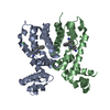
| ||||||||
|---|---|---|---|---|---|---|---|---|---|
| 1 |
| ||||||||
| Unit cell |
|
-Components
| #1: Protein | Mass: 23266.268 Da / Num. of mol.: 2 Source method: isolated from a genetically manipulated source Source: (gene. exp.) Streptomyces lividans (bacteria) / Strain: TK64 / Gene: ebrR / Plasmid: pET15b / Production host: #2: Chemical |   Mass: 58.693 Da / Num. of mol.: 2 / Source method: obtained synthetically / Formula: Ni Mass: 58.693 Da / Num. of mol.: 2 / Source method: obtained synthetically / Formula: Ni#3: Chemical |   Mass: 314.404 Da / Num. of mol.: 2 / Source method: obtained synthetically / Formula: C21H20N3 Mass: 314.404 Da / Num. of mol.: 2 / Source method: obtained synthetically / Formula: C21H20N3#4: Water | ChemComp-HOH / |  Mass: 18.015 Da / Num. of mol.: 24 / Source method: isolated from a natural source / Formula: H2O Mass: 18.015 Da / Num. of mol.: 24 / Source method: isolated from a natural source / Formula: H2O |
|---|
-Experimental details
-Experiment
| Experiment | Method: X-RAY DIFFRACTION / Number of used crystals: 1 |
|---|
- Sample preparation
| Crystal | Density Matthews: 2.94 Å3/Da / Density % sol: 58.17 % |
|---|---|
| Crystal grow | Temperature: 295 K / pH: 9 Details: 1.2 M Lithium sulfate, 0.1 M Tris-HCl, 0.01 M Nickel chloride, pH 9.0, VAPOR DIFFUSION, HANGING DROP, temperature 295K |
-Data collection
| Diffraction | Mean temperature: 100 K |
|---|---|
| Diffraction source | Source: SYNCHROTRON / Site: ALS  / Beamline: 8.3.1 / Wavelength: 1 / Beamline: 8.3.1 / Wavelength: 1 |
| Detector | Type: ADSC QUANTUM 315r / Detector: CCD / Date: Sep 27, 2008 |
| Radiation | Monochromator: SI(111) DOUBLE FLAT CRYSTAL / Protocol: SINGLE WAVELENGTH / Monochromatic (M) / Laue (L): M / Scattering type: x-ray |
| Radiation wavelength | Wavelength: 1 Å / Relative weight: 1 |
| Reflection | Resolution: 2.7→74.33 Å / Num. obs: 14305 / % possible obs: 98.1 % / Redundancy: 2.2 % / Rmerge(I) obs: 0.053 |
- Processing
| Software |
| ||||||||||||||||||||||||||||||||||||||||||||||||||||||||||||
|---|---|---|---|---|---|---|---|---|---|---|---|---|---|---|---|---|---|---|---|---|---|---|---|---|---|---|---|---|---|---|---|---|---|---|---|---|---|---|---|---|---|---|---|---|---|---|---|---|---|---|---|---|---|---|---|---|---|---|---|---|---|
| Refinement | Method to determine structure: MOLECULAR REPLACEMENT Starting model: PDB ENTRY 3HTA Resolution: 2.7→74.33 Å / Isotropic thermal model: ANISOTROPIC / Cross valid method: THROUGHOUT
| ||||||||||||||||||||||||||||||||||||||||||||||||||||||||||||
| Displacement parameters |
| ||||||||||||||||||||||||||||||||||||||||||||||||||||||||||||
| Refinement step | Cycle: LAST / Resolution: 2.7→74.33 Å
| ||||||||||||||||||||||||||||||||||||||||||||||||||||||||||||
| Refine LS restraints |
|
