 Movie
Movie Controller

[English] 日本語
 Yorodumi
- PDB-3hsu: Functional roles of the 6-s-cysteinyl, 8 alpha-N1-histidyl FAD in... -
+ Open data

- Basic information
| Entry | Database: PDB / ID: 3hsu | ||||||||||||
|---|---|---|---|---|---|---|---|---|---|---|---|---|---|
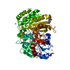
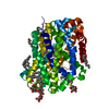
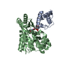
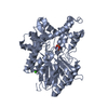
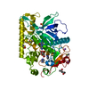
| Title | Functional roles of the 6-s-cysteinyl, 8 alpha-N1-histidyl FAD in Glucooligosaccharide Oxidase from Acremonium strictum | ||||||||||||
 Components Components | Glucooligosaccharide oxidase | ||||||||||||
 Keywords Keywords | OXIDOREDUCTASE / bicovalent flavoenzyme / (alpha + beta) / VAO family | ||||||||||||
| Function / homology |  Function and homology information Function and homology informationOxidoreductases; Acting on the CH-OH group of donors; With oxygen as acceptor / FAD binding / oxidoreductase activity / extracellular region / metal ion binding Similarity search - Function | ||||||||||||
| Biological species |  Acremonium strictum (fungus) Acremonium strictum (fungus) | ||||||||||||
| Method |  X-RAY DIFFRACTION / SYNCHROTRON / MOLECULAR REPLACEMENT / Resolution: 1.69 Å X-RAY DIFFRACTION / SYNCHROTRON / MOLECULAR REPLACEMENT / Resolution: 1.69 Å | ||||||||||||
 Authors Authors | Liaw, S.-H. / Huang, C.-H. | ||||||||||||
 Citation Citation | Journal: J.Biol.Chem. / Year: 2008 Title: Functional roles of the 6-S-cysteinyl, 8alpha-N1-histidyl FAD in glucooligosaccharide oxidase from Acremonium strictum Authors: Huang, C.-H. / Winkler, A. / Chen, C.-L. / Lai, W.-L. / Tsai, Y.-C. / Macheroux, P. / Liaw, S.-H. #1: Journal: J.Biol.Chem. / Year: 2005Title: Crystal structure of glucooligosaccharide oxidase from Acremonium strictum: a novel flavinylation of 6-S-cysteinyl, 8alpha-N1-histidyl FAD Authors: Huang, C.-H. / Lai, W.-L. / Lee, M.-H. / Chen, C.-J. / Vasella, A. / Tsai, Y.-C. / Liaw, S.-H. #2: Journal: Appl.Environ.Microbiol. / Year: 2005 Title: Structural characterization of glucooligosaccharide oxidase from Acremonium strictum Authors: Lee, M.-H. / Lai, W.-L. / Lin, S.-F. / Hsu, C.-S. / Liaw, S.-H. / Tsai, Y.-C. | ||||||||||||
| History |
|
- Structure visualization
| Structure viewer | Molecule: MolmilJmol/JSmol |
|---|
- Downloads & links
-Download
| PDBx/mmCIF format | 3hsu.cif.gz | 121.7 KB | Display | PDBx/mmCIF format |
|---|---|---|---|---|
| PDB format | pdb3hsu.ent.gz | 89.8 KB | Display | PDB format |
| PDBx/mmJSON format | 3hsu.json.gz | Tree view | PDBx/mmJSON format | |
| Others |  Other downloads Other downloads |
-Validation report
| Arichive directory | https://data.pdbj.org/pub/pdb/validation_reports/hs/3hsuftp://data.pdbj.org/pub/pdb/validation_reports/hs/3hsu | HTTPS FTP |
|---|
-Related structure data
| Related structure data |  1zr6S S: Starting model for refinement |
|---|---|
| Similar structure data |


-Links
 PDBj
PDBj

- Assembly
| Deposited unit | 
| ||||||||
|---|---|---|---|---|---|---|---|---|---|
| 1 |
| ||||||||
| Unit cell |
|
-Components
-Protein , 1 types, 1 molecules A
| #1: Protein | Mass: 55925.375 Da / Num. of mol.: 1 / Fragment: UNP residues 26-499 / Mutation: C130A Source method: isolated from a genetically manipulated source Source: (gene. exp.) Acremonium strictum (fungus) / Strain: T1 / Plasmid: pPICZ / Production host: Pichia pastoris (fungus) / Strain (production host): KM71References: UniProt: Q6PW77, Oxidoreductases; Acting on the CH-OH group of donors; With oxygen as acceptor |
|---|
-Sugars , 2 types, 2 molecules 
| #2: Polysaccharide | 2-acetamido-2-deoxy-beta-D-glucopyranose-(1-4)-2-acetamido-2-deoxy-beta-D-glucopyranose-(1-4)-2- ...2-acetamido-2-deoxy-beta-D-glucopyranose-(1-4)-2-acetamido-2-deoxy-beta-D-glucopyranose-(1-4)-2-acetamido-2-deoxy-beta-D-glucopyranose / triacetyl-beta-chitotriose  Source method: isolated from a genetically manipulated source Details: oligosaccharide / References: triacetyl-beta-chitotriose |
|---|---|
| #3: Sugar | ChemComp-NAG /  Type: D-saccharide, beta linking / Mass: 221.208 Da / Num. of mol.: 1 Type: D-saccharide, beta linking / Mass: 221.208 Da / Num. of mol.: 1Source method: isolated from a genetically manipulated source Formula: C8H15NO6 |
-Non-polymers , 3 types, 425 molecules 


| #4: Chemical | ChemComp-FAD /  Mass: 785.550 Da / Num. of mol.: 1 / Source method: obtained synthetically / Formula: C27H33N9O15P2 / Comment: FAD*YM Mass: 785.550 Da / Num. of mol.: 1 / Source method: obtained synthetically / Formula: C27H33N9O15P2 / Comment: FAD*YM |
|---|---|
| #5: Chemical | ChemComp-ZN /  Mass: 65.409 Da / Num. of mol.: 1 / Source method: obtained synthetically / Formula: Zn Mass: 65.409 Da / Num. of mol.: 1 / Source method: obtained synthetically / Formula: Zn |
| #6: Water | ChemComp-HOH / Mass: 18.015 Da / Num. of mol.: 423 / Source method: isolated from a natural source / Formula: H2O |
-Details
| Has protein modification | Y |
|---|
-Experimental details
-Experiment
| Experiment | Method: X-RAY DIFFRACTION / Number of used crystals: 1 |
|---|
- Sample preparation
| Crystal | Density Matthews: 2.5 Å3/Da / Density % sol: 50.9 % |
|---|---|
| Crystal grow | Temperature: 295 K / Method: vapor diffusion, hanging drop / pH: 6.5 Details: 25% PEG MME 550, 10mM ZnSO4, 0.1M MES, pH 6.5, VAPOR DIFFUSION, HANGING DROP, temperature 295K |
-Data collection
| Diffraction | Mean temperature: 100 K |
|---|---|
| Diffraction source | Source: SYNCHROTRON / Site: NSRRC  / Beamline: BL13B1 / Wavelength: 1 Å / Beamline: BL13B1 / Wavelength: 1 Å |
| Detector | Type: ADSC QUANTUM 315 / Detector: CCD / Date: Apr 24, 2008 |
| Radiation | Protocol: SINGLE WAVELENGTH / Monochromatic (M) / Laue (L): M / Scattering type: x-ray |
| Radiation wavelength | Wavelength: 1 Å / Relative weight: 1 |
| Reflection | Resolution: 1.69→50 Å / Num. all: 63644 / Num. obs: 62403 / % possible obs: 98 % / Observed criterion σ(F): 0 / Observed criterion σ(I): 0 / Redundancy: 13.5 % / Rmerge(I) obs: 0.061 / Rsym value: 0.061 / Net I/σ(I): 42.7 |
| Reflection shell | Resolution: 1.69→1.75 Å / Redundancy: 8.8 % / Rmerge(I) obs: 0.7 / Mean I/σ(I) obs: 2.2 / Num. unique all: 6225 / Rsym value: 0.73 / % possible all: 87 |
- Processing
| Software |
| ||||||||||||||||||||||||||||
|---|---|---|---|---|---|---|---|---|---|---|---|---|---|---|---|---|---|---|---|---|---|---|---|---|---|---|---|---|---|
| Refinement | Method to determine structure: MOLECULAR REPLACEMENT Starting model: PDB ENTRY 1ZR6 Resolution: 1.69→30.88 Å / Occupancy max: 1 / Occupancy min: 1 / Isotropic thermal model: isotropic B / Cross valid method: THROUGHOUT / σ(F): 0 / σ(I): 0 / Stereochemistry target values: Engh & Huber
| ||||||||||||||||||||||||||||
| Solvent computation | Bsol: 59.637 Å2 | ||||||||||||||||||||||||||||
| Displacement parameters | Biso max: 56.91 Å2 / Biso mean: 12.8 Å2 / Biso min: 12.97 Å2
| ||||||||||||||||||||||||||||
| Refine analyze | Luzzati coordinate error obs: 0.18 Å / Luzzati d res low obs: 6 Å / Luzzati sigma a obs: 0.15 Å | ||||||||||||||||||||||||||||
| Refinement step | Cycle: LAST / Resolution: 1.69→30.88 Å
| ||||||||||||||||||||||||||||
| Refine LS restraints |
| ||||||||||||||||||||||||||||
| LS refinement shell | Resolution: 1.69→1.7 Å
| ||||||||||||||||||||||||||||
| Xplor file |
|
