 Movie
Movie Controller

[English] 日本語
 Yorodumi
- PDB-3hoa: Crystal structure of the Thermus thermophilus M32 carboxypeptidase -
+ Open data

- Basic information
| Entry | Database: PDB / ID: 3hoa | ||||||
|---|---|---|---|---|---|---|---|
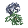
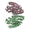
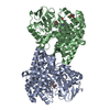
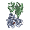
| Title | Crystal structure of the Thermus thermophilus M32 carboxypeptidase | ||||||
 Components Components | Thermostable carboxypeptidase 1 | ||||||
 Keywords Keywords | HYDROLASE / proline-rich loop / Carboxypeptidase | ||||||
| Function / homology |  Function and homology information Function and homology informationcarboxypeptidase Taq / metallocarboxypeptidase activity / proteolysis / zinc ion binding Similarity search - Function | ||||||
| Biological species |   Thermus thermophilus HB27 (bacteria) Thermus thermophilus HB27 (bacteria) | ||||||
| Method |  X-RAY DIFFRACTION / SYNCHROTRON / MOLECULAR REPLACEMENT / Resolution: 2.1 Å X-RAY DIFFRACTION / SYNCHROTRON / MOLECULAR REPLACEMENT / Resolution: 2.1 Å | ||||||
 Authors Authors | Lee, M.M. / Chan, M.K. | ||||||
 Citation Citation | Journal: Proteins / Year: 2009 Title: Insight into the substrate length restriction of M32 carboxypeptidases: Characterization of two distinct subfamilies. Authors: Lee, M.M. / Isaza, C.E. / White, J.D. / Chen, R.P. / Liang, G.F. / He, H.T. / Chan, S.I. / Chan, M.K. | ||||||
| History |
|
- Structure visualization
| Structure viewer | Molecule: MolmilJmol/JSmol |
|---|
- Downloads & links
-Download
| PDBx/mmCIF format | 3hoa.cif.gz | 216.3 KB | Display | PDBx/mmCIF format |
|---|---|---|---|---|
| PDB format | pdb3hoa.ent.gz | 173.3 KB | Display | PDB format |
| PDBx/mmJSON format | 3hoa.json.gz | Tree view | PDBx/mmJSON format | |
| Others |  Other downloads Other downloads |
-Validation report
| Arichive directory | https://data.pdbj.org/pub/pdb/validation_reports/ho/3hoaftp://data.pdbj.org/pub/pdb/validation_reports/ho/3hoa | HTTPS FTP |
|---|
-Related structure data


-Links
 PDBj
PDBj- Assembly
| Deposited unit | 
| ||||||||
|---|---|---|---|---|---|---|---|---|---|
| 1 |
| ||||||||
| Unit cell |
|
-Components
| #1: Protein | Mass: 58031.488 Da / Num. of mol.: 2 Source method: isolated from a genetically manipulated source Source: (gene. exp.) Thermus thermophilus HB27 (bacteria) / Strain: HB27 / DSM 7039 / Gene: TT_C1715 / Plasmid: pET28b / Production host: #2: Chemical | ChemComp-GOL / |   Mass: 92.094 Da / Num. of mol.: 1 / Source method: obtained synthetically / Formula: C3H8O3 Mass: 92.094 Da / Num. of mol.: 1 / Source method: obtained synthetically / Formula: C3H8O3#3: Water | ChemComp-HOH / |  Mass: 18.015 Da / Num. of mol.: 467 / Source method: isolated from a natural source / Formula: H2O Mass: 18.015 Da / Num. of mol.: 467 / Source method: isolated from a natural source / Formula: H2O |
|---|
-Experimental details
-Experiment
| Experiment | Method: X-RAY DIFFRACTION / Number of used crystals: 1 |
|---|
- Sample preparation
| Crystal | Density Matthews: 2.32 Å3/Da / Density % sol: 46.91 % |
|---|---|
| Crystal grow | Temperature: 298 K / Method: vapor diffusion, hanging drop / pH: 4 Details: 0.1 M Na-citrate pH 4.0, 8% w/v PEG 6000, VAPOR DIFFUSION, HANGING DROP, temperature 298K |
-Data collection
| Diffraction | Mean temperature: 100 K |
|---|---|
| Diffraction source | Source: SYNCHROTRON / Site: SSRL  / Beamline: BL1-5 / Wavelength: 1.00002 Å / Beamline: BL1-5 / Wavelength: 1.00002 Å |
| Detector | Type: ADSC QUANTUM 315 / Detector: CCD / Date: Sep 10, 2003 Details: 1m long Rh coated bent cylindrical mirror for horizontal and vertical focusing |
| Radiation | Monochromator: Si(111) / Protocol: SINGLE WAVELENGTH / Monochromatic (M) / Laue (L): M / Scattering type: x-ray |
| Radiation wavelength | Wavelength: 1.00002 Å / Relative weight: 1 |
| Reflection | Resolution: 2.1→45.7 Å / Num. all: 60029 / Num. obs: 60029 / % possible obs: 99.9 % / Observed criterion σ(F): 0 / Observed criterion σ(I): 0 / Redundancy: 3.417 % / Biso Wilson estimate: 2.7 Å2 / Rmerge(I) obs: 0.095 / Net I/σ(I): 20.4 |
| Reflection shell | Resolution: 2.1→2.23 Å / Rmerge(I) obs: 0.209 / Mean I/σ(I) obs: 5.1 / Num. unique all: 8712 / % possible all: 99.9 |
- Processing
| Software |
| ||||||||||||||||||||||||||||||||||||
|---|---|---|---|---|---|---|---|---|---|---|---|---|---|---|---|---|---|---|---|---|---|---|---|---|---|---|---|---|---|---|---|---|---|---|---|---|---|
| Refinement | Method to determine structure: MOLECULAR REPLACEMENT Starting model: Bacillus subtilis carboxypeptidase Resolution: 2.1→19.98 Å / Rfactor Rfree error: 0.003 / Data cutoff high absF: 132243.15 / Data cutoff low absF: 0 / Isotropic thermal model: RESTRAINED / Cross valid method: THROUGHOUT / σ(F): 0 / σ(I): 0 / Stereochemistry target values: Engh & Huber
| ||||||||||||||||||||||||||||||||||||
| Solvent computation | Solvent model: FLAT MODEL / Bsol: 43.7339 Å2 / ksol: 0.380191 e/Å3 | ||||||||||||||||||||||||||||||||||||
| Displacement parameters | Biso mean: 20.6 Å2
| ||||||||||||||||||||||||||||||||||||
| Refine analyze |
| ||||||||||||||||||||||||||||||||||||
| Refinement step | Cycle: LAST / Resolution: 2.1→19.98 Å
| ||||||||||||||||||||||||||||||||||||
| Refine LS restraints |
| ||||||||||||||||||||||||||||||||||||
| LS refinement shell | Resolution: 2.1→2.23 Å / Rfactor Rfree error: 0.009 / Total num. of bins used: 6
| ||||||||||||||||||||||||||||||||||||
| Xplor file |
|
