 Movie
Movie Controller

[English] 日本語
 Yorodumi
- PDB-3hii: Crystal structure of human diamine oxidase in complex with the in... -
+ Open data

- Basic information
| Entry | Database: PDB / ID: 3hii | |||||||||
|---|---|---|---|---|---|---|---|---|---|---|
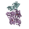
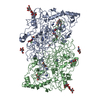
| Title | Crystal structure of human diamine oxidase in complex with the inhibitor pentamidine | |||||||||
 Components Components | Amiloride-sensitive amine oxidase | |||||||||
 Keywords Keywords | OXIDOREDUCTASE / copper amine oxidase / topaquinone / TPQ / diamine oxidase / DAO / human / pentamidine / Glycoprotein / Heparin-binding / Metal-binding / Secreted | |||||||||
| Function / homology |  Function and homology information Function and homology informationdiamine oxidase / putrescine metabolic process / histamine oxidase activity / putrescine oxidase activity / diamine oxidase activity / primary methylamine oxidase activity / Phase I - Functionalization of compounds / bicellular tight junction / quinone binding / specific granule lumen ...diamine oxidase / putrescine metabolic process / histamine oxidase activity / putrescine oxidase activity / diamine oxidase activity / primary methylamine oxidase activity / Phase I - Functionalization of compounds / bicellular tight junction / quinone binding / specific granule lumen / peroxisome / heparin binding / copper ion binding / calcium ion binding / Neutrophil degranulation / protein homodimerization activity / : / extracellular exosome / extracellular region / plasma membrane Similarity search - Function | |||||||||
| Biological species |  Homo sapiens (human) Homo sapiens (human) | |||||||||
| Method |  X-RAY DIFFRACTION / BY REFINEMENT / Resolution: 2.149 Å X-RAY DIFFRACTION / BY REFINEMENT / Resolution: 2.149 Å | |||||||||
 Authors Authors | McGrath, A.P. / Guss, J.M. | |||||||||
 Citation Citation | Journal: Biochemistry / Year: 2009 Title: Structure and inhibition of human diamine oxidase Authors: McGrath, A.P. / Hilmer, K.M. / Collyer, C.A. / Shepard, E.M. / Elmore, B.O. / Brown, D.E. / Dooley, D.M. / Guss, J.M. | |||||||||
| History |
|
- Structure visualization
| Structure viewer | Molecule: MolmilJmol/JSmol |
|---|
- Downloads & links
-Download
| PDBx/mmCIF format | 3hii.cif.gz | 326.9 KB | Display | PDBx/mmCIF format |
|---|---|---|---|---|
| PDB format | pdb3hii.ent.gz | 258.5 KB | Display | PDB format |
| PDBx/mmJSON format | 3hii.json.gz | Tree view | PDBx/mmJSON format | |
| Others |  Other downloads Other downloads |
-Validation report
| Arichive directory | https://data.pdbj.org/pub/pdb/validation_reports/hi/3hiiftp://data.pdbj.org/pub/pdb/validation_reports/hi/3hii | HTTPS FTP |
|---|
-Related structure data
| Related structure data |  3hi7SC  3higC S: Starting model for refinement C: citing same article ( |
|---|---|
| Similar structure data |


-Links
 PDBj
PDBj- Assembly
| Deposited unit | 
| ||||||||
|---|---|---|---|---|---|---|---|---|---|
| 1 |
| ||||||||
| Unit cell |
|
-Components
-Protein , 1 types, 2 molecules AB
| #1: Protein | Mass: 83463.797 Da / Num. of mol.: 2 Source method: isolated from a genetically manipulated source Source: (gene. exp.) Homo sapiens (human) / Gene: ABP1 / Cell line (production host): Schneider 2 / Production host:  |
|---|
-Sugars , 2 types, 6 molecules
| #2: Polysaccharide | beta-D-mannopyranose-(1-4)-2-acetamido-2-deoxy-beta-D-glucopyranose-(1-4)-2-acetamido-2-deoxy-beta- ...beta-D-mannopyranose-(1-4)-2-acetamido-2-deoxy-beta-D-glucopyranose-(1-4)-2-acetamido-2-deoxy-beta-D-glucopyranose Source method: isolated from a genetically manipulated source |
|---|---|
| #3: Polysaccharide | 2-acetamido-2-deoxy-beta-D-glucopyranose-(1-4)-2-acetamido-2-deoxy-beta-D-glucopyranose Source method: isolated from a genetically manipulated source |
-Non-polymers , 5 types, 925 molecules 


| #4: Chemical |  Mass: 63.546 Da / Num. of mol.: 2 / Source method: obtained synthetically / Formula: Cu Mass: 63.546 Da / Num. of mol.: 2 / Source method: obtained synthetically / Formula: Cu#5: Chemical | ChemComp-CA /  Mass: 40.078 Da / Num. of mol.: 4 / Source method: obtained synthetically / Formula: Ca Mass: 40.078 Da / Num. of mol.: 4 / Source method: obtained synthetically / Formula: Ca#6: Chemical | ChemComp-GOL / |  Mass: 92.094 Da / Num. of mol.: 1 / Source method: obtained synthetically / Formula: C3H8O3 Mass: 92.094 Da / Num. of mol.: 1 / Source method: obtained synthetically / Formula: C3H8O3#7: Chemical |  Mass: 340.419 Da / Num. of mol.: 2 / Source method: obtained synthetically / Formula: C19H24N4O2 / Comment: medication, Antimicrobial*YM Mass: 340.419 Da / Num. of mol.: 2 / Source method: obtained synthetically / Formula: C19H24N4O2 / Comment: medication, Antimicrobial*YM#8: Water | ChemComp-HOH / | Mass: 18.015 Da / Num. of mol.: 916 / Source method: isolated from a natural source / Formula: H2O |
|---|
-Experimental details
-Experiment
| Experiment | Method: X-RAY DIFFRACTION / Number of used crystals: 1 |
|---|
- Sample preparation
| Crystal | Density Matthews: 2.58 Å3/Da / Density % sol: 52.24 % / Mosaicity: 0.431 ° |
|---|---|
| Crystal grow | Temperature: 298 K / Method: vapor diffusion, hanging drop / pH: 7.5 Details: 0.1M bis-tris propane, 20%(w/v) PEG 3350, 0.2M sodium sulfate, pH 7.5, vapor diffusion, hanging drop, temperature 298K |
-Data collection
| Diffraction | Mean temperature: 100 K |
|---|---|
| Diffraction source | Source: ROTATING ANODE / Type: RIGAKU RU200 / Wavelength: 1.5418 Å |
| Detector | Type: MAR scanner 345 mm plate / Detector: IMAGE PLATE / Date: Nov 5, 2008 / Details: OSMIC MIRRORS |
| Radiation | Monochromator: Ni FILTER / Protocol: SINGLE WAVELENGTH / Monochromatic (M) / Laue (L): M / Scattering type: x-ray |
| Radiation wavelength | Wavelength: 1.5418 Å / Relative weight: 1 |
| Reflection | Resolution: 2.149→50 Å / Num. all: 89751 / Num. obs: 89751 / % possible obs: 95 % / Observed criterion σ(F): 0 / Observed criterion σ(I): 0 / Redundancy: 3 % / Biso Wilson estimate: 29.96 Å2 / Rmerge(I) obs: 0.103 / Χ2: 1.067 / Net I/σ(I): 9.903 |
| Reflection shell | Resolution: 2.149→2.23 Å / Redundancy: 2.4 % / Rmerge(I) obs: 0.336 / Mean I/σ(I) obs: 2.7 / Num. unique all: 8090 / Χ2: 1.041 / % possible all: 87.1 |
- Processing
| Software |
| ||||||||||||||||||||||||||||||||||||||||||||||||||||||||||||||||||||||||||||||||||||||||||||||||||||||||||||||||||||||||||||||||||||||||||||||||||||||||||||||||||||||||||
|---|---|---|---|---|---|---|---|---|---|---|---|---|---|---|---|---|---|---|---|---|---|---|---|---|---|---|---|---|---|---|---|---|---|---|---|---|---|---|---|---|---|---|---|---|---|---|---|---|---|---|---|---|---|---|---|---|---|---|---|---|---|---|---|---|---|---|---|---|---|---|---|---|---|---|---|---|---|---|---|---|---|---|---|---|---|---|---|---|---|---|---|---|---|---|---|---|---|---|---|---|---|---|---|---|---|---|---|---|---|---|---|---|---|---|---|---|---|---|---|---|---|---|---|---|---|---|---|---|---|---|---|---|---|---|---|---|---|---|---|---|---|---|---|---|---|---|---|---|---|---|---|---|---|---|---|---|---|---|---|---|---|---|---|---|---|---|---|---|---|---|---|
| Refinement | Method to determine structure: BY REFINEMENT Starting model: PDB ENTRY 3HI7 Resolution: 2.149→25.84 Å / Cor.coef. Fo:Fc: 0.948 / Cor.coef. Fo:Fc free: 0.926 / Occupancy max: 1 / Occupancy min: 0.3 / FOM work R set: 0.866 / SU B: 4.494 / SU ML: 0.118 / SU R Cruickshank DPI: 0.245 / SU Rfree: 0.19 / Cross valid method: THROUGHOUT / σ(F): 0 / ESU R: 0.245 / ESU R Free: 0.19 / Stereochemistry target values: MAXIMUM LIKELIHOOD Details: HYDROGENS HAVE BEEN ADDED IN THE RIDING POSITIONS U VALUES: REFINED INDIVIDUALLY
| ||||||||||||||||||||||||||||||||||||||||||||||||||||||||||||||||||||||||||||||||||||||||||||||||||||||||||||||||||||||||||||||||||||||||||||||||||||||||||||||||||||||||||
| Solvent computation | Ion probe radii: 0.8 Å / Shrinkage radii: 0.8 Å / VDW probe radii: 1.2 Å / Solvent model: MASK | ||||||||||||||||||||||||||||||||||||||||||||||||||||||||||||||||||||||||||||||||||||||||||||||||||||||||||||||||||||||||||||||||||||||||||||||||||||||||||||||||||||||||||
| Displacement parameters | Biso max: 62.23 Å2 / Biso mean: 21.017 Å2 / Biso min: 11.86 Å2
| ||||||||||||||||||||||||||||||||||||||||||||||||||||||||||||||||||||||||||||||||||||||||||||||||||||||||||||||||||||||||||||||||||||||||||||||||||||||||||||||||||||||||||
| Refinement step | Cycle: LAST / Resolution: 2.149→25.84 Å
| ||||||||||||||||||||||||||||||||||||||||||||||||||||||||||||||||||||||||||||||||||||||||||||||||||||||||||||||||||||||||||||||||||||||||||||||||||||||||||||||||||||||||||
| Refine LS restraints |
| ||||||||||||||||||||||||||||||||||||||||||||||||||||||||||||||||||||||||||||||||||||||||||||||||||||||||||||||||||||||||||||||||||||||||||||||||||||||||||||||||||||||||||
| LS refinement shell | Resolution: 2.149→2.204 Å / Total num. of bins used: 20
|
