[English] 日本語
Yorodumi
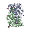
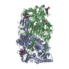
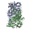
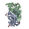
- PDB-2y73: THE NATIVE STRUCTURES OF SOLUBLE HUMAN PRIMARY AMINE OXIDASE AOC3 -

+
Open data


ID or keywords:

Loading...

-
Basic information

Entry
Database: PDB / ID: 2y73
TitleTHE NATIVE STRUCTURES OF SOLUBLE HUMAN PRIMARY AMINE OXIDASE AOC3
ComponentsMEMBRANE PRIMARY AMINE OXIDASE
KeywordsOXIDOREDUCTASE
Function / homology
Function and homology information


primary-amine oxidase / aliphatic amine oxidase activity / primary methylamine oxidase activity / amine metabolic process / Phase I - Functionalization of compounds / microvillus / quinone binding / early endosome / cell adhesion / inflammatory response ...primary-amine oxidase / aliphatic amine oxidase activity / primary methylamine oxidase activity / amine metabolic process / Phase I - Functionalization of compounds / microvillus / quinone binding / early endosome / cell adhesion / inflammatory response / copper ion binding / protein heterodimerization activity / response to antibiotic / calcium ion binding / Golgi apparatus / cell surface / endoplasmic reticulum / identical protein binding / membrane / plasma membrane / cytoplasm
Similarity search - Function
Copper amine oxidase, N3-terminal / Copper amine oxidase, N2-terminal / Copper amine oxidase, N2 domain / Copper amine oxidase, N3 domain / Copper amine oxidase, catalytic domain / Copper amine oxidase copper-binding site signature. / Copper amine oxidase topaquinone signature. / Nuclear Transport Factor 2; Chain: A, - #40 / Copper amine oxidase / Copper amine oxidase, catalytic domain ...Copper amine oxidase, N3-terminal / Copper amine oxidase, N2-terminal / Copper amine oxidase, N2 domain / Copper amine oxidase, N3 domain / Copper amine oxidase, catalytic domain / Copper amine oxidase copper-binding site signature. / Copper amine oxidase topaquinone signature. / Nuclear Transport Factor 2; Chain: A, - #40 / Copper amine oxidase / Copper amine oxidase, catalytic domain / Copper amine oxidase, N-terminal / Copper amine oxidase, catalytic domain superfamily / Copper amine oxidase, enzyme domain / Beta-galactosidase; Chain A, domain 5 / Nuclear Transport Factor 2; Chain: A, / Distorted Sandwich / Roll / Mainly Beta / Alpha Beta
Similarity search - Domain/homology
COPPER (II) ION / FORMIC ACID / IMIDAZOLE / Amine oxidase [copper-containing] 3
Similarity search - Component
Biological speciesHOMO SAPIENS (human)
MethodX-RAY DIFFRACTION / SYNCHROTRON / MOLECULAR REPLACEMENT / Resolution: 2.6 Å
AuthorsElovaara, H. / Kidron, H. / Parkash, V. / Nymalm, Y. / Bligt, E. / Ollikka, P. / Smith, D.J. / Pihlavisto, M. / Salmi, M. / Jalkanen, S. / Salminen, T.A.
CitationJournal: Biochemistry / Year: 2011
Title: Identification of Two Imidazole Binding Sites and Key Residues for Substrate Specificity in Human Primary Amine Oxidase Aoc3.
Authors: Elovaara, H. / Kidron, H. / Parkash, V. / Nymalm, Y. / Bligt, E. / Ollikka, P. / Smith, D.J. / Pihlavisto, M. / Salmi, M. / Jalkanen, S. / Salminen, T.A.
History
DepositionJan 28, 2011Deposition site: PDBE / Processing site: PDBE
Revision 1.0Jun 15, 2011Provider: repository / Type: Initial release
Revision 1.1Aug 10, 2011Group: Database references / Version format compliance
Revision 1.2Jul 12, 2017Group: Data collection / Category: diffrn_source / Item: _diffrn_source.pdbx_synchrotron_site
Revision 2.0Jul 29, 2020Group: Atomic model / Data collection ...Atomic model / Data collection / Derived calculations / Other / Structure summary
Category: atom_site / chem_comp ...atom_site / chem_comp / entity / pdbx_branch_scheme / pdbx_chem_comp_identifier / pdbx_database_status / pdbx_entity_branch / pdbx_entity_branch_descriptor / pdbx_entity_branch_link / pdbx_entity_branch_list / pdbx_entity_nonpoly / pdbx_nonpoly_scheme / pdbx_struct_assembly_gen / pdbx_struct_conn_angle / struct_asym / struct_conn / struct_site / struct_site_gen
Item: _atom_site.B_iso_or_equiv / _atom_site.Cartn_x ..._atom_site.B_iso_or_equiv / _atom_site.Cartn_x / _atom_site.Cartn_y / _atom_site.Cartn_z / _atom_site.auth_asym_id / _atom_site.auth_atom_id / _atom_site.auth_comp_id / _atom_site.auth_seq_id / _atom_site.label_asym_id / _atom_site.label_atom_id / _atom_site.label_comp_id / _atom_site.label_entity_id / _atom_site.type_symbol / _chem_comp.name / _chem_comp.type / _pdbx_database_status.status_code_sf / _pdbx_struct_assembly_gen.asym_id_list / _pdbx_struct_conn_angle.ptnr1_label_asym_id / _pdbx_struct_conn_angle.ptnr2_label_asym_id / _pdbx_struct_conn_angle.ptnr3_label_asym_id / _struct_conn.conn_type_id / _struct_conn.id / _struct_conn.pdbx_dist_value / _struct_conn.pdbx_leaving_atom_flag / _struct_conn.pdbx_role / _struct_conn.ptnr1_auth_asym_id / _struct_conn.ptnr1_auth_comp_id / _struct_conn.ptnr1_auth_seq_id / _struct_conn.ptnr1_label_asym_id / _struct_conn.ptnr1_label_atom_id / _struct_conn.ptnr1_label_comp_id / _struct_conn.ptnr1_label_seq_id / _struct_conn.ptnr2_auth_asym_id / _struct_conn.ptnr2_auth_comp_id / _struct_conn.ptnr2_auth_seq_id / _struct_conn.ptnr2_label_asym_id / _struct_conn.ptnr2_label_atom_id / _struct_conn.ptnr2_label_comp_id / _struct_conn.ptnr2_label_seq_id
Description: Carbohydrate remediation / Provider: repository / Type: Remediation
Revision 2.1Dec 20, 2023Group: Data collection / Database references ...Data collection / Database references / Refinement description / Structure summary
Category: chem_comp / chem_comp_atom ...chem_comp / chem_comp_atom / chem_comp_bond / database_2 / pdbx_initial_refinement_model
Item: _chem_comp.pdbx_synonyms / _database_2.pdbx_DOI / _database_2.pdbx_database_accession

-
Structure visualization

Structure viewerMolecule:
MolmilJmol/JSmol

Downloads & links

-
Assembly

Deposited unit
A: MEMBRANE PRIMARY AMINE OXIDASE
B: MEMBRANE PRIMARY AMINE OXIDASE
hetero molecules


Theoretical massNumber of molelcules
Total (without water)173,30826
Polymers169,4692
Non-polymers3,83924
Water6,233346
1


  • Idetical with deposited unit
  • defined by author&software
TypeNameSymmetry operationNumber
identity operation1_555x,y,z1
Buried area20250 Å2
ΔGint-126.3 kcal/mol
Surface area46190 Å2
MethodPISA
Unit cell
Length a, b, c (Å)225.755, 225.755, 216.985
Angle α, β, γ (deg.)90.00, 90.00, 120.00
Int Tables number179
Space group name H-MP6522

-
Components

-
Protein , 1 types, 2 molecules AB

#1: Protein MEMBRANE PRIMARY AMINE OXIDASE / AMINE OXIDASE COPPER CONTAINING 3 / COPPER AMINE OXIDASE / HPAO / SEMICARBAZIDE-SENSITIVE AMINE ...AMINE OXIDASE COPPER CONTAINING 3 / COPPER AMINE OXIDASE / HPAO / SEMICARBAZIDE-SENSITIVE AMINE OXIDASE / SSAO / VASCULAR ADHESION PROTEIN 1 / VAP-1


Mass: 84734.562 Da / Num. of mol.: 2 / Source method: isolated from a natural source / Source: (natural) HOMO SAPIENS (human) / References: UniProt: Q16853, primary-amine oxidase

-
Sugars , 3 types, 8 molecules

#2: Polysaccharide alpha-D-mannopyranose-(1-4)-2-acetamido-2-deoxy-beta-D-glucopyranose-(1-4)-2-acetamido-2-deoxy-beta- ...alpha-D-mannopyranose-(1-4)-2-acetamido-2-deoxy-beta-D-glucopyranose-(1-4)-2-acetamido-2-deoxy-beta-D-glucopyranose


Type: oligosaccharide / Mass: 586.542 Da / Num. of mol.: 2
Source method: isolated from a genetically manipulated source
DescriptorTypeProgram
DManpa1-4DGlcpNAcb1-4DGlcpNAcb1-Glycam Condensed SequenceGMML 1.0
WURCS=2.0/2,3,2/[a2122h-1b_1-5_2*NCC/3=O][a1122h-1a_1-5]/1-1-2/a4-b1_b4-c1WURCSPDB2Glycan 1.1.0
[]{[(4+1)][b-D-GlcpNAc]{[(4+1)][b-D-GlcpNAc]{[(4+1)][a-D-Manp]{}}}}LINUCSPDB-CARE
#3: Polysaccharide 2-acetamido-2-deoxy-beta-D-glucopyranose-(1-4)-2-acetamido-2-deoxy-beta-D-glucopyranose


Type: oligosaccharide / Mass: 424.401 Da / Num. of mol.: 2
Source method: isolated from a genetically manipulated source
DescriptorTypeProgram
DGlcpNAcb1-4DGlcpNAcb1-Glycam Condensed SequenceGMML 1.0
WURCS=2.0/1,2,1/[a2122h-1b_1-5_2*NCC/3=O]/1-1/a4-b1WURCSPDB2Glycan 1.1.0
[]{[(4+1)][b-D-GlcpNAc]{[(4+1)][b-D-GlcpNAc]{}}}LINUCSPDB-CARE
#7: Sugar
ChemComp-NAG / 2-acetamido-2-deoxy-beta-D-glucopyranose / N-acetyl-beta-D-glucosamine / 2-acetamido-2-deoxy-beta-D-glucose / 2-acetamido-2-deoxy-D-glucose / 2-acetamido-2-deoxy-glucose / N-ACETYL-D-GLUCOSAMINE


Type: D-saccharide, beta linking / Mass: 221.208 Da / Num. of mol.: 4
Source method: isolated from a genetically manipulated source
Formula: C8H15NO6
IdentifierTypeProgram
DGlcpNAcbCONDENSED IUPAC CARBOHYDRATE SYMBOLGMML 1.0
N-acetyl-b-D-glucopyranosamineCOMMON NAMEGMML 1.0
b-D-GlcpNAcIUPAC CARBOHYDRATE SYMBOLPDB-CARE 1.0
GlcNAcSNFG CARBOHYDRATE SYMBOLGMML 1.0

-
Non-polymers , 5 types, 362 molecules

#4: Chemical ChemComp-CU / COPPER (II) ION


Mass: 63.546 Da / Num. of mol.: 2 / Source method: obtained synthetically / Formula: Cu
#5: Chemical
ChemComp-CA / CALCIUM ION


Mass: 40.078 Da / Num. of mol.: 4 / Source method: obtained synthetically / Formula: Ca
#6: Chemical
ChemComp-IMD / IMIDAZOLE


Mass: 69.085 Da / Num. of mol.: 8 / Source method: obtained synthetically / Formula: C3H5N2
#8: Chemical ChemComp-FMT / FORMIC ACID


Mass: 46.025 Da / Num. of mol.: 2 / Source method: obtained synthetically / Formula: CH2O2
#9: Water ChemComp-HOH / water


Mass: 18.015 Da / Num. of mol.: 346 / Source method: isolated from a natural source / Formula: H2O

-
Experimental details

-
Experiment

ExperimentMethod: X-RAY DIFFRACTION / Number of used crystals: 1

-
Sample preparation

CrystalDensity Matthews: 2.2 Å3/Da / Density % sol: 45 % / Description: NONE
Crystal growpH: 7.4
Details: 0.7 M POTASSIUM/SODIUM TARTRATE, 100 MM IMIDAZOLE PH7.4, 200 MM NACL, 2M SODIUM FORMATE

-
Data collection

DiffractionMean temperature: 183 K
Diffraction sourceSource: SYNCHROTRON / Site: MAX II / Beamline: I711 / Wavelength: 0.9059
DetectorType: MARRESEARCH SX-165 / Detector: CCD / Date: Jun 9, 2006 / Details: VERTICALLY FOCUSING CYLINDRICAL MIRROR
RadiationMonochromator: SINGLE ASYMMETRICALLY CUT SI(111) CRYSTAL WITH HORIZONTAL DIFFRACTION PLANE
Protocol: SINGLE WAVELENGTH / Monochromatic (M) / Laue (L): M / Scattering type: x-ray
Radiation wavelengthWavelength: 0.9059 Å / Relative weight: 1
ReflectionResolution: 2.6→29.93 Å / Num. obs: 99745 / % possible obs: 99.9 % / Observed criterion σ(I): 3 / Redundancy: 11.8 % / Biso Wilson estimate: 36.03 Å2 / Rmerge(I) obs: 0.01 / Net I/σ(I): 17.5
Reflection shellResolution: 2.6→2.7 Å / Redundancy: 11.9 % / Rmerge(I) obs: 0.05 / Mean I/σ(I) obs: 5.1 / % possible all: 100

-
Processing

Software
NameVersionClassification
PHENIX(PHENIX.REFINE)refinement
XDSdata reduction
XSCALEdata scaling
PHASERphasing
RefinementMethod to determine structure: MOLECULAR REPLACEMENT
Starting model: PDB ENTRY 1US1
Resolution: 2.6→19.931 Å / SU ML: 0.32 / σ(F): 1.38 / Phase error: 20.91 / Stereochemistry target values: ML
RfactorNum. reflection% reflection
Rfree0.2126 4978 5 %
Rwork0.1748 --
obs0.1767 99551 99.99 %
Solvent computationShrinkage radii: 0.95 Å / VDW probe radii: 1.2 Å / Solvent model: FLAT BULK SOLVENT MODEL / Bsol: 20.63 Å2 / ksol: 0.327 e/Å3
Displacement parametersBiso mean: 37.3 Å2
Baniso -1Baniso -2Baniso -3
1--0.7569 Å20 Å20 Å2
2---0.7569 Å20 Å2
3---1.5137 Å2
Refinement stepCycle: LAST / Resolution: 2.6→19.931 Å
ProteinNucleic acidLigandSolventTotal
Num. atoms11087 0 242 346 11675
Refine LS restraints
Refine-IDTypeDev idealNumber
X-RAY DIFFRACTIONf_bond_d0.01611695
X-RAY DIFFRACTIONf_angle_d1.77615949
X-RAY DIFFRACTIONf_dihedral_angle_d25.9514389
X-RAY DIFFRACTIONf_chiral_restr0.1031704
X-RAY DIFFRACTIONf_plane_restr0.0122082
LS refinement shell
Resolution (Å)Rfactor RfreeNum. reflection RfreeRfactor RworkNum. reflection RworkRefine-ID% reflection obs (%)
2.6-2.69270.34264910.29969337X-RAY DIFFRACTION100
2.6927-2.80030.31014900.27149312X-RAY DIFFRACTION100
2.8003-2.92730.29684930.25729368X-RAY DIFFRACTION100
2.9273-3.08120.28844920.24399340X-RAY DIFFRACTION100
3.0812-3.27340.2574940.20549387X-RAY DIFFRACTION100
3.2734-3.52490.2274950.17739407X-RAY DIFFRACTION100
3.5249-3.87720.19884970.14939437X-RAY DIFFRACTION100
3.8772-4.43290.15684990.12149485X-RAY DIFFRACTION100
4.4329-5.56480.14135050.119586X-RAY DIFFRACTION100
5.5648-19.93120.17155220.15669914X-RAY DIFFRACTION100

+
About Yorodumi

-
News

-
Feb 9, 2022. New format data for meta-information of EMDB entries

New format data for meta-information of EMDB entries

  • Version 3 of the EMDB header file is now the official format.
  • The previous official version 1.9 will be removed from the archive.

Related info.:EMDB header

External links:wwPDB to switch to version 3 of the EMDB data model

-
Aug 12, 2020. Covid-19 info

Covid-19 info

URL: https://pdbjlvh1.pdbj.org/emnavi/covid19.php

New page: Covid-19 featured information page in EM Navigator.

Related info.:Covid-19 info / Mar 5, 2020. Novel coronavirus structure data

+
Mar 5, 2020. Novel coronavirus structure data

Novel coronavirus structure data

Related info.:Yorodumi Speices / Aug 12, 2020. Covid-19 info

External links:COVID-19 featured content - PDBj / Molecule of the Month (242):Coronavirus Proteases

+
Jan 31, 2019. EMDB accession codes are about to change! (news from PDBe EMDB page)

EMDB accession codes are about to change! (news from PDBe EMDB page)

  • The allocation of 4 digits for EMDB accession codes will soon come to an end. Whilst these codes will remain in use, new EMDB accession codes will include an additional digit and will expand incrementally as the available range of codes is exhausted. The current 4-digit format prefixed with “EMD-” (i.e. EMD-XXXX) will advance to a 5-digit format (i.e. EMD-XXXXX), and so on. It is currently estimated that the 4-digit codes will be depleted around Spring 2019, at which point the 5-digit format will come into force.
  • The EM Navigator/Yorodumi systems omit the EMD- prefix.

Related info.:Q: What is EMD? / ID/Accession-code notation in Yorodumi/EM Navigator

External links:EMDB Accession Codes are Changing Soon! / Contact to PDBj

+
Jul 12, 2017. Major update of PDB

Major update of PDB

  • wwPDB released updated PDB data conforming to the new PDBx/mmCIF dictionary.
  • This is a major update changing the version number from 4 to 5, and with Remediation, in which all the entries are updated.
  • In this update, many items about electron microscopy experimental information are reorganized (e.g. em_software).
  • Now, EM Navigator and Yorodumi are based on the updated data.

External links:wwPDB Remediation / Enriched Model Files Conforming to OneDep Data Standards Now Available in the PDB FTP Archive

-
Yorodumi

Thousand views of thousand structures

  • Yorodumi is a browser for structure data from EMDB, PDB, SASBDB, etc.
  • This page is also the successor to EM Navigator detail page, and also detail information page/front-end page for Omokage search.
  • The word "yorodu" (or yorozu) is an old Japanese word meaning "ten thousand". "mi" (miru) is to see.

Related info.:EMDB / PDB / SASBDB / Comparison of 3 databanks / Yorodumi Search / Aug 31, 2016. New EM Navigator & Yorodumi / Yorodumi Papers / Jmol/JSmol / Function and homology information / Changes in new EM Navigator and Yorodumi

Read more