 Movie
Movie Controller

[English] 日本語
 Yorodumi
- PDB-3hhn: Crystal structure of class I ligase ribozyme self-ligation produc... -
+ Open data

- Basic information
| Entry | Database: PDB / ID: 3hhn | ||||||
|---|---|---|---|---|---|---|---|
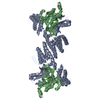
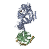
| Title | Crystal structure of class I ligase ribozyme self-ligation product, in complex with U1A RBD | ||||||
 Components Components |
| ||||||
 Keywords Keywords | LIGASE/RNA / Ligase Ribozyme / Ribozyme / Catalytic RNA / Protein-RNA complex / Ribonucleoprotein / RNA-binding / LIGASE-RNA COMPLEX | ||||||
| Function / homology |  Function and homology information Function and homology informationU1 snRNP binding / U1 snRNP / U1 snRNA binding / U4/U6 x U5 tri-snRNP complex / mRNA Splicing - Major Pathway / spliceosomal complex / mRNA splicing, via spliceosome / DNA binding / RNA binding / nucleoplasm ...U1 snRNP binding / U1 snRNP / U1 snRNA binding / U4/U6 x U5 tri-snRNP complex / mRNA Splicing - Major Pathway / spliceosomal complex / mRNA splicing, via spliceosome / DNA binding / RNA binding / nucleoplasm / identical protein binding / nucleus Similarity search - Function | ||||||
| Biological species |  Homo sapiens (human) Homo sapiens (human) | ||||||
| Method |  X-RAY DIFFRACTION / SYNCHROTRON / SIRAS / Resolution: 2.987 Å X-RAY DIFFRACTION / SYNCHROTRON / SIRAS / Resolution: 2.987 Å | ||||||
 Authors Authors | Shechner, D.M. / Grant, R.A. / Bagby, S.C. / Bartel, D.P. | ||||||
 Citation Citation | Journal: Science / Year: 2009 Title: Crystal structure of the catalytic core of an RNA-polymerase ribozyme. Authors: Shechner, D.M. / Grant, R.A. / Bagby, S.C. / Koldobskaya, Y. / Piccirilli, J.A. / Bartel, D.P. | ||||||
| History |
|
- Structure visualization
| Structure viewer | Molecule: MolmilJmol/JSmol |
|---|
- Downloads & links
-Download
| PDBx/mmCIF format | 3hhn.cif.gz | 214.5 KB | Display | PDBx/mmCIF format |
|---|---|---|---|---|
| PDB format | pdb3hhn.ent.gz | 160.3 KB | Display | PDB format |
| PDBx/mmJSON format | 3hhn.json.gz | Tree view | PDBx/mmJSON format | |
| Others |  Other downloads Other downloads |
-Validation report
| Arichive directory | https://data.pdbj.org/pub/pdb/validation_reports/hh/3hhnftp://data.pdbj.org/pub/pdb/validation_reports/hh/3hhn | HTTPS FTP |
|---|
-Related structure data


-Links
 PDBj
PDBj


- Assembly
| Deposited unit | 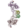
| ||||||||||||||||||||
|---|---|---|---|---|---|---|---|---|---|---|---|---|---|---|---|---|---|---|---|---|---|
| 1 |
| ||||||||||||||||||||
| Unit cell |
| ||||||||||||||||||||
| Noncrystallographic symmetry (NCS) | NCS oper:
| ||||||||||||||||||||
| Details | THIS CRYSTAL STRUCTURE IS NOT A BIOLOGICAL ASSEMBLY. IN SOLUTION, THE RNA-U1A COMPLEX IS EXPECTED TO HAVE ONE COPY OF EACH MOLECULE. |
-Components
| #1: Protein | Mass: 11209.120 Da / Num. of mol.: 2 / Fragment: RNA binding domain: UNP residues 1-100 / Mutation: Y31H, Q31R Source method: isolated from a genetically manipulated source Details: Overexpressed in E. coli from a pET11a derivative bearing the the "double" mutant U1A RBD (1-98) Source: (gene. exp.) Homo sapiens (human) / Gene: SNRPA / Plasmid: Modified pET11a / Production host:  #2: RNA chain | Mass: 44168.270 Da / Num. of mol.: 2 / Source method: obtained synthetically Details: Synthetic construct transcribed in vitro using T7 RNA polymerase off of a linearized plasmid pUC307HP #3: Chemical | ChemComp-MG /   Mass: 24.305 Da / Num. of mol.: 34 / Source method: obtained synthetically / Formula: Mg Mass: 24.305 Da / Num. of mol.: 34 / Source method: obtained synthetically / Formula: Mg#4: Water | ChemComp-HOH / |  Mass: 18.015 Da / Num. of mol.: 192 / Source method: isolated from a natural source / Formula: H2O Mass: 18.015 Da / Num. of mol.: 192 / Source method: isolated from a natural source / Formula: H2O |
|---|
-Experimental details
-Experiment
| Experiment | Method: X-RAY DIFFRACTION / Number of used crystals: 1 |
|---|
- Sample preparation
| Crystal | Density Matthews: 2.49 Å3/Da / Density % sol: 50.58 % | ||||||||||||||||||||||||||||||||||||
|---|---|---|---|---|---|---|---|---|---|---|---|---|---|---|---|---|---|---|---|---|---|---|---|---|---|---|---|---|---|---|---|---|---|---|---|---|---|
| Crystal grow | Temperature: 293 K / Method: vapor diffusion, hanging drop / pH: 6 Details: 50mM Sodium cacodylate pH 6.0, 40mM Magnesium acetate, 26% 2-methyl-2,4-pentane diol (MPD), 1mM Spermine HCl. Diffraction-quality crystals grown by microseeding., VAPOR DIFFUSION, HANGING DROP, temperature 293K | ||||||||||||||||||||||||||||||||||||
| Components of the solutions |
|
-Data collection
| Diffraction | Mean temperature: 100 K | ||||||||||||||||||||||||||||||||||||||||||||||||||||||||||||||||||
|---|---|---|---|---|---|---|---|---|---|---|---|---|---|---|---|---|---|---|---|---|---|---|---|---|---|---|---|---|---|---|---|---|---|---|---|---|---|---|---|---|---|---|---|---|---|---|---|---|---|---|---|---|---|---|---|---|---|---|---|---|---|---|---|---|---|---|---|
| Diffraction source | Source: SYNCHROTRON / Site: APS  / Beamline: 8-BM / Wavelength: 0.9792 Å / Beamline: 8-BM / Wavelength: 0.9792 Å | ||||||||||||||||||||||||||||||||||||||||||||||||||||||||||||||||||
| Detector | Type: ADSC QUANTUM 315 / Detector: CCD / Date: Jul 22, 2004 | ||||||||||||||||||||||||||||||||||||||||||||||||||||||||||||||||||
| Radiation | Monochromator: Crystal / Protocol: SINGLE WAVELENGTH / Monochromatic (M) / Laue (L): M / Scattering type: x-ray | ||||||||||||||||||||||||||||||||||||||||||||||||||||||||||||||||||
| Radiation wavelength | Wavelength: 0.9792 Å / Relative weight: 1 | ||||||||||||||||||||||||||||||||||||||||||||||||||||||||||||||||||
| Reflection | Resolution: 2.987→50 Å / Num. obs: 21075 / % possible obs: 97 % / Observed criterion σ(F): 0 / Observed criterion σ(I): 0 / Redundancy: 3.7 % / Rmerge(I) obs: 0.069 / Χ2: 2.471 / Net I/σ(I): 28.185 | ||||||||||||||||||||||||||||||||||||||||||||||||||||||||||||||||||
| Reflection shell |
|
- Processing
| Software |
| ||||||||||||||||||||||||||||||||||||||||||||||||||||||||||||||||||||||||||||||||||||||||||||||||||||||||||||||||||||||||||||||||||||||||||||||||||||||||||||||||||||||||||||||||||||||||||||||||||||||||||||||||||||||||||||||||||||||||||||||||||||||
|---|---|---|---|---|---|---|---|---|---|---|---|---|---|---|---|---|---|---|---|---|---|---|---|---|---|---|---|---|---|---|---|---|---|---|---|---|---|---|---|---|---|---|---|---|---|---|---|---|---|---|---|---|---|---|---|---|---|---|---|---|---|---|---|---|---|---|---|---|---|---|---|---|---|---|---|---|---|---|---|---|---|---|---|---|---|---|---|---|---|---|---|---|---|---|---|---|---|---|---|---|---|---|---|---|---|---|---|---|---|---|---|---|---|---|---|---|---|---|---|---|---|---|---|---|---|---|---|---|---|---|---|---|---|---|---|---|---|---|---|---|---|---|---|---|---|---|---|---|---|---|---|---|---|---|---|---|---|---|---|---|---|---|---|---|---|---|---|---|---|---|---|---|---|---|---|---|---|---|---|---|---|---|---|---|---|---|---|---|---|---|---|---|---|---|---|---|---|---|---|---|---|---|---|---|---|---|---|---|---|---|---|---|---|---|---|---|---|---|---|---|---|---|---|---|---|---|---|---|---|---|---|---|---|---|---|---|---|---|---|---|---|---|---|---|---|---|---|
| Refinement | Method to determine structure: SIRAS / Resolution: 2.987→40.121 Å / Occupancy max: 1 / Occupancy min: 0.5 / FOM work R set: 0.773 / σ(F): 0 / σ(I): 0 / Stereochemistry target values: MAXIMUM LIKELIHOOD
| ||||||||||||||||||||||||||||||||||||||||||||||||||||||||||||||||||||||||||||||||||||||||||||||||||||||||||||||||||||||||||||||||||||||||||||||||||||||||||||||||||||||||||||||||||||||||||||||||||||||||||||||||||||||||||||||||||||||||||||||||||||||
| Solvent computation | Bsol: 29.083 Å2 / ksol: 0.249 e/Å3 | ||||||||||||||||||||||||||||||||||||||||||||||||||||||||||||||||||||||||||||||||||||||||||||||||||||||||||||||||||||||||||||||||||||||||||||||||||||||||||||||||||||||||||||||||||||||||||||||||||||||||||||||||||||||||||||||||||||||||||||||||||||||
| Displacement parameters | Biso max: 174.15 Å2 / Biso mean: 77.482 Å2 / Biso min: 30.91 Å2
| ||||||||||||||||||||||||||||||||||||||||||||||||||||||||||||||||||||||||||||||||||||||||||||||||||||||||||||||||||||||||||||||||||||||||||||||||||||||||||||||||||||||||||||||||||||||||||||||||||||||||||||||||||||||||||||||||||||||||||||||||||||||
| Refine analyze | Luzzati coordinate error obs: 0.33 Å | ||||||||||||||||||||||||||||||||||||||||||||||||||||||||||||||||||||||||||||||||||||||||||||||||||||||||||||||||||||||||||||||||||||||||||||||||||||||||||||||||||||||||||||||||||||||||||||||||||||||||||||||||||||||||||||||||||||||||||||||||||||||
| Refinement step | Cycle: LAST / Resolution: 2.987→40.121 Å
| ||||||||||||||||||||||||||||||||||||||||||||||||||||||||||||||||||||||||||||||||||||||||||||||||||||||||||||||||||||||||||||||||||||||||||||||||||||||||||||||||||||||||||||||||||||||||||||||||||||||||||||||||||||||||||||||||||||||||||||||||||||||
| Refine LS restraints |
| ||||||||||||||||||||||||||||||||||||||||||||||||||||||||||||||||||||||||||||||||||||||||||||||||||||||||||||||||||||||||||||||||||||||||||||||||||||||||||||||||||||||||||||||||||||||||||||||||||||||||||||||||||||||||||||||||||||||||||||||||||||||
| LS refinement shell |
|
