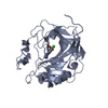
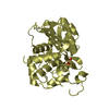
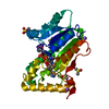
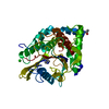
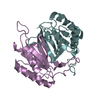
- PDB-3h51: Crystal structure of Putative calcium/calmodulin dependent protei... -
+
Open data
ID or keywords:
Loading...
-
Basic information
Entry
Database: PDB / ID: 3h51
Title
Crystal structure of Putative calcium/calmodulin dependent protein kinase II association domain (NP_636218.1) from XANTHOMONAS CAMPESTRIS at 1.70 A resolution
Components
Putative calcium/calmodulin dependent protein kinase II association domain
Keywords
PROTEIN BINDING / NP_636218.1 / Putative calcium/calmodulin dependent protein kinase II association domain / Structural Genomics / Joint Center for Structural Genomics / JCSG / Protein Structure Initiative / PSI-2 / unknown function
Uncharacterised conserved protein UCP028470, steroid isomerase-related / Steroid delta5-4-isomerase / Calcium/calmodulin-dependent protein kinase II, association-domain / Calcium/calmodulin dependent protein kinase II association domain / Nuclear Transport Factor 2; Chain: A, - #50 / NTF2-like domain superfamily / Nuclear Transport Factor 2; Chain: A, / Roll / Alpha Beta Similarity search - Domain/homology
DI(HYDROXYETHYL)ETHER / Unknown ligand / Calcium/calmodulin-dependent protein kinase II association-domain domain-containing protein Similarity search - Component
Biological species
Xanthomonas campestris pv. campestris (bacteria)
Method
X-RAY DIFFRACTION / SYNCHROTRON / MAD / Resolution: 1.7 Å
#191 - Nov 2015 Glutamate-gated Chloride Receptors similarity (1)
-
Assembly
Deposited unit
A: Putative calcium/calmodulin dependent protein kinase II association domain B: Putative calcium/calmodulin dependent protein kinase II association domain hetero molecules
Mass: 18.015 Da / Num. of mol.: 251 / Source method: isolated from a natural source / Formula: H2O
Has protein modification
Y
Sequence details
THE CONSTRUCT (RESIDUES 20-174) WAS EXPRESSED WITH A PURIFICATION TAG MGSDKIHHHHHHENLYFQG. THE TAG ...THE CONSTRUCT (RESIDUES 20-174) WAS EXPRESSED WITH A PURIFICATION TAG MGSDKIHHHHHHENLYFQG. THE TAG WAS REMOVED WITH TEV PROTEASE LEAVING ONLY A GLYCINE (0) FOLLOWED BY THE TARGET SEQUENCE.
-
Experimental details
-
Experiment
Experiment
Method: X-RAY DIFFRACTION / Number of used crystals: 1
-
Sample preparation
Crystal
Density Matthews: 2.32 Å3/Da / Density % sol: 46.96 %
Crystal grow
Temperature: 277 K / Method: vapor diffusion, sitting drop / pH: 8.5 Details: 20.0000% PEG-1000, 0.1M TRIS pH 8.5, NANODROP, VAPOR DIFFUSION, SITTING DROP, temperature 277K
Type: MARMOSAIC 325 mm CCD / Detector: CCD / Date: Feb 18, 2009 / Details: Flat mirror (vertical focusing)
Radiation
Monochromator: Single crystal Si(111) bent monochromator (horizontal focusing) Protocol: MAD / Monochromatic (M) / Laue (L): M / Scattering type: x-ray
Radiation wavelength
ID
Wavelength (Å)
Relative weight
1
0.91837
1
2
0.97901
1
Reflection
Resolution: 1.7→29.488 Å / Num. obs: 33865 / % possible obs: 97.9 % / Redundancy: 3.2 % / Biso Wilson estimate: 22.186 Å2 / Rmerge(I) obs: 0.074 / Rsym value: 0.074 / Net I/σ(I): 6.464
Reflection shell
Diffraction-ID: 1
Resolution (Å)
Redundancy (%)
Rmerge(I) obs
Mean I/σ(I) obs
Num. measured all
Num. unique all
Rsym value
% possible all
1.7-1.74
3.2
0.665
1.2
7906
2496
0.665
97.3
1.74-1.79
3.2
0.525
1.5
7586
2398
0.525
97.4
1.79-1.84
3.2
0.372
2.1
7475
2355
0.372
96.5
1.84-1.9
3.2
0.278
2.8
7228
2285
0.278
98.5
1.9-1.96
3.2
0.231
3.1
7022
2213
0.231
96.9
1.96-2.03
3.2
0.188
4
6846
2158
0.188
97.4
2.03-2.11
3.2
0.158
4.7
6574
2085
0.158
98.6
2.11-2.19
3.2
0.135
5.3
6314
1990
0.135
97.6
2.19-2.29
3.2
0.113
5.5
6106
1931
0.113
97.4
2.29-2.4
3.1
0.098
6.6
5831
1854
0.098
98.8
2.4-2.53
3.2
0.085
7.8
5603
1760
0.085
98.6
2.53-2.69
3.2
0.076
8.7
5253
1662
0.076
98.3
2.69-2.87
3.2
0.07
9.1
4983
1570
0.07
98.3
2.87-3.1
3.2
0.066
9.4
4566
1443
0.066
98.6
3.1-3.4
3.1
0.055
11.7
4300
1367
0.055
98.8
3.4-3.8
3.1
0.047
12.7
3802
1208
0.047
98.7
3.8-4.39
3.1
0.044
13.1
3427
1088
0.044
98.2
4.39-5.38
3.1
0.049
11.9
2837
910
0.049
99.1
5.38-7.6
3.1
0.056
10.5
2199
713
0.056
98.2
7.6-29.49
3.1
0.047
13.3
1157
379
0.047
94.4
-
Phasing
Phasing
Method: MAD
-
Processing
Software
Name
Version
Classification
NB
REFMAC
5.2.0019
refinement
PHENIX
refinement
SHELX
phasing
MolProbity
3beta29
modelbuilding
SCALA
3.2.5
datascaling
PDB_EXTRACT
3.006
dataextraction
MOSFLM
datareduction
autoSHARP
phasing
Refinement
Method to determine structure: MAD / Resolution: 1.7→29.488 Å / Cor.coef. Fo:Fc: 0.961 / Cor.coef. Fo:Fc free: 0.946 / Occupancy max: 1 / Occupancy min: 0.25 / SU B: 4.425 / SU ML: 0.075 / TLS residual ADP flag: LIKELY RESIDUAL / Cross valid method: THROUGHOUT / σ(F): 0 / ESU R: 0.108 / ESU R Free: 0.107 Stereochemistry target values: MAXIMUM LIKELIHOOD WITH PHASES Details: 1. HYDROGENS HAVE BEEN ADDED IN THE RIDING POSITIONS. 2. ATOM RECORDS CONTAIN RESIDUAL B FACTORS ONLY. 3. A MET-INHIBITION PROTOCOL WAS USED FOR SELENOMETHIONINE INCORPORATION DURING PROTEIN ...Details: 1. HYDROGENS HAVE BEEN ADDED IN THE RIDING POSITIONS. 2. ATOM RECORDS CONTAIN RESIDUAL B FACTORS ONLY. 3. A MET-INHIBITION PROTOCOL WAS USED FOR SELENOMETHIONINE INCORPORATION DURING PROTEIN EXPRESSION. THE OCCUPANCY OF THE SE ATOMS IN THE MSE RESIDUES WAS REDUCED TO 0.75 FOR THE REDUCED SCATTERING POWER DUE TO PARTIAL S-MET INCORPORATION. 4. PEG AND PG4 MOLECULES (FRAGMENTS OF PEG1000 AND PEG200) USED IN THE CRYSTALLIZATION AND CRYOPROTECTION CONDITION HAVE BEEN MODELED. 5. ELECTRON DENSITIES OF RESIDUES OF 30-34 AND 108-112 IN BOTH A AND B CHAINS ARE POOR AND MODELS ARE NOT RELIABLE IN THIS REGION. 6. AN UNIDENTIFIED LIGAND (UNL) HAS BEEN MODELED AT THE PUTATIVE ACTIVE SITE OF BOTH A AND B CHAINS.
Rfactor
Num. reflection
% reflection
Selection details
Rfree
0.215
1695
5 %
RANDOM
Rwork
0.179
-
-
-
obs
0.18
33842
97.62 %
-
Solvent computation
Ion probe radii: 0.8 Å / Shrinkage radii: 0.8 Å / VDW probe radii: 1.2 Å / Solvent model: MASK
In the structure databanks used in Yorodumi, some data are registered as the other names, "COVID-19 virus" and "2019-nCoV". Here are the details of the virus and the list of structure data.
Jan 31, 2019. EMDB accession codes are about to change! (news from PDBe EMDB page)
EMDB accession codes are about to change! (news from PDBe EMDB page)
The allocation of 4 digits for EMDB accession codes will soon come to an end. Whilst these codes will remain in use, new EMDB accession codes will include an additional digit and will expand incrementally as the available range of codes is exhausted. The current 4-digit format prefixed with “EMD-” (i.e. EMD-XXXX) will advance to a 5-digit format (i.e. EMD-XXXXX), and so on. It is currently estimated that the 4-digit codes will be depleted around Spring 2019, at which point the 5-digit format will come into force.
The EM Navigator/Yorodumi systems omit the EMD- prefix.
Related info.:Q: What is EMD? / ID/Accession-code notation in Yorodumi/EM Navigator
Yorodumi is a browser for structure data from EMDB, PDB, SASBDB, etc.
This page is also the successor to EM Navigator detail page, and also detail information page/front-end page for Omokage search.
The word "yorodu" (or yorozu) is an old Japanese word meaning "ten thousand". "mi" (miru) is to see.
Related info.:EMDB / PDB / SASBDB / Comparison of 3 databanks / Yorodumi Search / Aug 31, 2016. New EM Navigator & Yorodumi / Yorodumi Papers / Jmol/JSmol / Function and homology information / Changes in new EM Navigator and Yorodumi
 Movie
Movie Controller

 Yorodumi
 Open data
 Basic information
 Components
 Keywords
 Function and homology information
 Xanthomonas campestris pv. campestris (bacteria)
 X-RAY DIFFRACTION /
  Authors
 Citation
 Structure visualization
 Downloads & links
 Other downloads


 PDBj
PDBj


 Assembly

 Mass: 106.120 Da / Num. of mol.: 2 / Source method: obtained synthetically / Formula: C4H10O3
 Mass: 194.226 Da / Num. of mol.: 1 / Source method: obtained synthetically / Formula: C8H18O5 / Comment: precipitant*YM
 Mass: 18.015 Da / Num. of mol.: 251 / Source method: isolated from a natural source / Formula: H2O
 Sample preparation
 / Beamline: BL11-1 / Wavelength: 0.91837,0.97901
 Processing