 Movie
Movie Controller

[English] 日本語
 Yorodumi
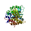
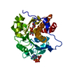
- PDB-3h4w: Structure of a Ca+2 dependent Phosphatidylinositol-specific phosp... -
+ Open data

- Basic information
| Entry | Database: PDB / ID: 3h4w | ||||||
|---|---|---|---|---|---|---|---|
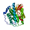
| Title | Structure of a Ca+2 dependent Phosphatidylinositol-specific phospholipase C (PI-PLC) Enzyme from Streptomyces antibioticus | ||||||
 Components Components | Phosphatidylinositol-specific phospholipase C1 | ||||||
 Keywords Keywords | HYDROLASE / PI-PLC / Ca2+-dependent / catalytic TIM barrel / disulfide-linked helix-loop | ||||||
| Function / homology |  Function and homology information Function and homology informationphosphoinositide phospholipase C / phosphatidylinositol-4,5-bisphosphate phospholipase C activity / lipid metabolic process Similarity search - Function | ||||||
| Biological species |  Streptomyces antibioticus (bacteria) Streptomyces antibioticus (bacteria) | ||||||
| Method |  X-RAY DIFFRACTION / SYNCHROTRON / SAD / Resolution: 1.5 Å X-RAY DIFFRACTION / SYNCHROTRON / SAD / Resolution: 1.5 Å | ||||||
 Authors Authors | Jackson, M.R. / Selby, T.L. | ||||||
 Citation Citation | Journal: To be Published Title: Crystal Structure of a Ca2+-dependent PI-PLC Authors: Jackson, M.R. / Selby, T.L. | ||||||
| History |
|
- Structure visualization
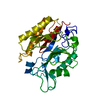
| Structure viewer | Molecule: MolmilJmol/JSmol |
|---|
- Downloads & links
-Download
| PDBx/mmCIF format | 3h4w.cif.gz | 144.3 KB | Display | PDBx/mmCIF format |
|---|---|---|---|---|
| PDB format | pdb3h4w.ent.gz | 111.2 KB | Display | PDB format |
| PDBx/mmJSON format | 3h4w.json.gz | Tree view | PDBx/mmJSON format | |
| Others |  Other downloads Other downloads |
-Validation report
| Arichive directory | https://data.pdbj.org/pub/pdb/validation_reports/h4/3h4wftp://data.pdbj.org/pub/pdb/validation_reports/h4/3h4w | HTTPS FTP |
|---|
-Related structure data


-Links
 PDBj
PDBj


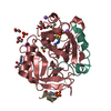
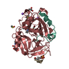
- Assembly
| Deposited unit | 
| ||||||||
|---|---|---|---|---|---|---|---|---|---|
| 1 |
| ||||||||
| Unit cell |
| ||||||||
| Components on special symmetry positions |
|
-Components
-Protein , 1 types, 1 molecules A
| #1: Protein | Mass: 36867.422 Da / Num. of mol.: 1 Source method: isolated from a genetically manipulated source Details: The N-terminal aa sequence in the gene (1.mngkrcvgtasraaaaviavsgpaga.26) is cleaved off by the organism and was replaced with the HIS-tagged sequence (1.MGSSHHHHHHSSGLVPRGSHM.21) for protein ...Details: The N-terminal aa sequence in the gene (1.mngkrcvgtasraaaaviavsgpaga.26) is cleaved off by the organism and was replaced with the HIS-tagged sequence (1.MGSSHHHHHHSSGLVPRGSHM.21) for protein expression. Selenomethionine incorporation using auxotrophic strain and synthetically substituted media. Source: (gene. exp.) Streptomyces antibioticus (bacteria) / Gene: plc1, plc1 AB439135.1:1234..2268 / Plasmid: pET28b / Production host: References: UniProt: B3A043, phosphoinositide phospholipase C |
|---|
-Non-polymers , 5 types, 328 molecules 


| #2: Chemical | ChemComp-CL /  Mass: 35.453 Da / Num. of mol.: 1 / Source method: obtained synthetically / Formula: Cl Mass: 35.453 Da / Num. of mol.: 1 / Source method: obtained synthetically / Formula: Cl | ||||||
|---|---|---|---|---|---|---|---|
| #3: Chemical | ChemComp-EOH /  Mass: 46.068 Da / Num. of mol.: 8 / Source method: obtained synthetically / Formula: C2H6O Mass: 46.068 Da / Num. of mol.: 8 / Source method: obtained synthetically / Formula: C2H6O#4: Chemical |  Mass: 44.053 Da / Num. of mol.: 2 / Source method: obtained synthetically / Formula: C2H4O Mass: 44.053 Da / Num. of mol.: 2 / Source method: obtained synthetically / Formula: C2H4O#5: Chemical |  Mass: 92.094 Da / Num. of mol.: 3 / Source method: obtained synthetically / Formula: C3H8O3 Mass: 92.094 Da / Num. of mol.: 3 / Source method: obtained synthetically / Formula: C3H8O3#6: Water | ChemComp-HOH / | Mass: 18.015 Da / Num. of mol.: 314 / Source method: isolated from a natural source / Formula: H2O |
-Details
| Has protein modification | Y |
|---|---|
| Sequence details | THE RESIDUE 264 IN COORDINATE |
-Experimental details
-Experiment
| Experiment | Method: X-RAY DIFFRACTION / Number of used crystals: 1 |
|---|
- Sample preparation
| Crystal | Density Matthews: 2.28 Å3/Da / Density % sol: 46.07 % |
|---|---|
| Crystal grow | Temperature: 277 K / Method: vapor diffusion, hanging drop / pH: 4.6 Details: 21% PEG 10000, 85mM sodium acetate pH 4.6, 170mM ammonium acetate, 9% glycerol. SeMe-derived crystals obtained with initial microseeding with underivatized crystals followed by a series of ...Details: 21% PEG 10000, 85mM sodium acetate pH 4.6, 170mM ammonium acetate, 9% glycerol. SeMe-derived crystals obtained with initial microseeding with underivatized crystals followed by a series of macroseedings with derivatized enzyme and same conditions., VAPOR DIFFUSION, HANGING DROP, temperature 277K |
-Data collection
| Diffraction | Mean temperature: 100 K |
|---|---|
| Diffraction source | Source: SYNCHROTRON / Site: SSRL  / Beamline: BL9-2 / Wavelength: 0.9796 Å / Beamline: BL9-2 / Wavelength: 0.9796 Å |
| Detector | Type: MARMOSAIC 325 mm CCD / Detector: CCD / Date: Jan 1, 2008 |
| Radiation | Protocol: SAD / Scattering type: x-ray |
| Radiation wavelength | Wavelength: 0.9796 Å / Relative weight: 1 |
| Reflection | Resolution: 1.5→77.61 Å / Num. all: 52173 / Num. obs: 52171 / % possible obs: 95.15 % / Observed criterion σ(I): 3.7 / Redundancy: 11.65 % / Biso Wilson estimate: 13.6 Å2 / Rmerge(I) obs: 0.058 / Net I/σ(I): 8.2689 |
| Reflection shell | Resolution: 1.5→1.58 Å / Redundancy: 5.76 % / Rmerge(I) obs: 0.22 / Mean I/σ(I) obs: 3.19 / Num. measured all: 36537 / Num. unique all: 6345 / % possible all: 81.66 |
-Phasing
| Phasing | Method: SAD | ||||||||||||||||||||||||||||||||||||||||||||||||||||||||||||||||||||||||||||||||||||
|---|---|---|---|---|---|---|---|---|---|---|---|---|---|---|---|---|---|---|---|---|---|---|---|---|---|---|---|---|---|---|---|---|---|---|---|---|---|---|---|---|---|---|---|---|---|---|---|---|---|---|---|---|---|---|---|---|---|---|---|---|---|---|---|---|---|---|---|---|---|---|---|---|---|---|---|---|---|---|---|---|---|---|---|---|---|
| Phasing MAD set site |
|
- Processing
| Software |
| |||||||||||||||||||||||||||||||||||||||||||||||||||||||||||||||||||||||||||||||||||||||||||||||||||||||||||||||||||||||||||||||||||||||||||||||||||
|---|---|---|---|---|---|---|---|---|---|---|---|---|---|---|---|---|---|---|---|---|---|---|---|---|---|---|---|---|---|---|---|---|---|---|---|---|---|---|---|---|---|---|---|---|---|---|---|---|---|---|---|---|---|---|---|---|---|---|---|---|---|---|---|---|---|---|---|---|---|---|---|---|---|---|---|---|---|---|---|---|---|---|---|---|---|---|---|---|---|---|---|---|---|---|---|---|---|---|---|---|---|---|---|---|---|---|---|---|---|---|---|---|---|---|---|---|---|---|---|---|---|---|---|---|---|---|---|---|---|---|---|---|---|---|---|---|---|---|---|---|---|---|---|---|---|---|---|---|
| Refinement | Method to determine structure: SAD / Resolution: 1.5→77.59 Å / Cor.coef. Fo:Fc: 0.947 / Cor.coef. Fo:Fc free: 0.944 / WRfactor Rfree: 0.211 / WRfactor Rwork: 0.192 / Occupancy max: 1 / Occupancy min: 0.5 / SU B: 2.281 / SU ML: 0.04 / ESU R: 0.103 / ESU R Free: 0.077 / Stereochemistry target values: MAXIMUM LIKELIHOO Details: HYDROGENS HAVE BEEN ADDED IN THE RIDING POSITIONS U VALUES : REFINED INDIVIDUALLY
| |||||||||||||||||||||||||||||||||||||||||||||||||||||||||||||||||||||||||||||||||||||||||||||||||||||||||||||||||||||||||||||||||||||||||||||||||||
| Solvent computation | Ion probe radii: 0.8 Å / Shrinkage radii: 0.8 Å / VDW probe radii: 1.2 Å / Solvent model: BABINET MODEL WITH MASK | |||||||||||||||||||||||||||||||||||||||||||||||||||||||||||||||||||||||||||||||||||||||||||||||||||||||||||||||||||||||||||||||||||||||||||||||||||
| Displacement parameters | Biso max: 54.16 Å2 / Biso mean: 14.295 Å2 / Biso min: 6.39 Å2
| |||||||||||||||||||||||||||||||||||||||||||||||||||||||||||||||||||||||||||||||||||||||||||||||||||||||||||||||||||||||||||||||||||||||||||||||||||
| Refinement step | Cycle: LAST / Resolution: 1.5→77.59 Å
| |||||||||||||||||||||||||||||||||||||||||||||||||||||||||||||||||||||||||||||||||||||||||||||||||||||||||||||||||||||||||||||||||||||||||||||||||||
| Refine LS restraints |
| |||||||||||||||||||||||||||||||||||||||||||||||||||||||||||||||||||||||||||||||||||||||||||||||||||||||||||||||||||||||||||||||||||||||||||||||||||
| LS refinement shell | Refine-ID: X-RAY DIFFRACTION / Total num. of bins used: 20
|
