Resolution: 1.78→1.84 Å / Redundancy: 3.3 % / Rmerge(I) obs: 0.254 / Mean I/σ(I) obs: 4.6 / Num. unique all: 8596 / % possible all: 78.4
-
Processing
Software
Name
Version
Classification
NB
REFMAC
5.2.0019
refinement
PDB_EXTRACT
3
dataextraction
HKL-2000
datacollection
DENZO
datareduction
SCALEPACK
datascaling
SnB
phasing
RESOLVE
phasing
Refinement
Method to determine structure: SAD / Resolution: 1.78→50 Å / Cor.coef. Fo:Fc: 0.954 / Cor.coef. Fo:Fc free: 0.922 / SU B: 5.365 / SU ML: 0.086 / Cross valid method: THROUGHOUT / σ(F): 0 / ESU R: 0.232 / ESU R Free: 0.142 / Stereochemistry target values: MAXIMUM LIKELIHOOD / Details: The Friedel pairs were used in phasing
Rfactor
Num. reflection
% reflection
Selection details
Rfree
0.254
2131
5 %
RANDOM
Rwork
0.193
-
-
-
obs
0.196
42277
93.76 %
-
Solvent computation
Ion probe radii: 0.8 Å / Shrinkage radii: 0.8 Å / VDW probe radii: 1.2 Å / Solvent model: MASK
Displacement parameters
Biso mean: 17.052 Å2
Baniso -1
Baniso -2
Baniso -3
1-
0.07 Å2
0 Å2
0 Å2
2-
-
-0.02 Å2
0 Å2
3-
-
-
-0.05 Å2
Refinement step
Cycle: LAST / Resolution: 1.78→50 Å
Protein
Nucleic acid
Ligand
Solvent
Total
Num. atoms
3233
0
96
360
3689
Refine LS restraints
Refine-ID
Type
Dev ideal
Dev ideal target
Number
X-RAY DIFFRACTION
r_bond_refined_d
0.008
0.022
3407
X-RAY DIFFRACTION
r_angle_refined_deg
1.187
1.998
4671
X-RAY DIFFRACTION
r_dihedral_angle_1_deg
6.013
5
430
X-RAY DIFFRACTION
r_dihedral_angle_2_deg
35.769
23.676
136
X-RAY DIFFRACTION
r_dihedral_angle_3_deg
14.705
15
497
X-RAY DIFFRACTION
r_dihedral_angle_4_deg
21.559
15
22
X-RAY DIFFRACTION
r_chiral_restr
0.082
0.2
524
X-RAY DIFFRACTION
r_gen_planes_refined
0.014
0.02
2560
X-RAY DIFFRACTION
r_nbd_refined
0.225
0.2
1627
X-RAY DIFFRACTION
r_nbtor_refined
0.321
0.2
2313
X-RAY DIFFRACTION
r_xyhbond_nbd_refined
0.157
0.2
318
X-RAY DIFFRACTION
r_symmetry_vdw_refined
0.233
0.2
52
X-RAY DIFFRACTION
r_symmetry_hbond_refined
0.127
0.2
10
X-RAY DIFFRACTION
r_mcbond_it
3.285
1.5
2194
X-RAY DIFFRACTION
r_mcangle_it
4.243
2
3430
X-RAY DIFFRACTION
r_scbond_it
7.091
3
1359
X-RAY DIFFRACTION
r_scangle_it
9.229
4.5
1241
LS refinement shell
Resolution: 1.78→1.82 Å / Total num. of bins used: 20
Rfactor
Num. reflection
% reflection
Rfree
0.284
120
-
Rwork
0.204
2366
-
obs
-
2486
76.59 %
Refinement TLS params.
Method: refined / Refine-ID: X-RAY DIFFRACTION
ID
L11 (°2)
L12 (°2)
L13 (°2)
L22 (°2)
L23 (°2)
L33 (°2)
S11 (Å °)
S12 (Å °)
S13 (Å °)
S21 (Å °)
S22 (Å °)
S23 (Å °)
S31 (Å °)
S32 (Å °)
S33 (Å °)
T11 (Å2)
T12 (Å2)
T13 (Å2)
T22 (Å2)
T23 (Å2)
T33 (Å2)
Origin x (Å)
Origin y (Å)
Origin z (Å)
1
0.1151
-0.0276
-0.0489
0.1605
-0.0575
0.1262
-0.0082
-0.0045
0.0121
0.005
0.0013
0.0069
-0.0096
-0.0111
0.0068
-0.0088
-0.0019
-0.0021
-0.0044
-0.0032
-0.0031
19.679
50.304
42.676
2
0.191
-0.0369
-0.0114
0.1856
0.083
0.1973
-0.0091
0.0141
0.0029
-0.0056
0.0004
-0.0123
-0.0163
0.0132
0.0087
-0.0114
-0.0029
-0.0026
-0.0061
-0.0006
-0.0042
28.026
50.173
11.872
3
4.6015
-4.0075
1.0346
4.9455
-0.4188
0.3924
0.0878
0.0367
-0.0428
-0.163
-0.1302
0.0404
-0.0587
0.0478
0.0423
-0.0102
-0.0176
-0.0023
-0.0092
-0.0045
-0.0378
26.95
46.098
45.476
4
2.7204
3.3783
1.4155
4.8798
1.5633
0.7919
0.0437
0.0039
-0.0522
0.1683
-0.0017
0.0943
0.0037
0.0374
-0.0419
-0.0071
-0.0002
0.0127
-0.0016
0.0104
-0.0129
20.701
46.051
9.231
Refinement TLS group
ID
Refine-ID
Refine TLS-ID
Auth asym-ID
Auth seq-ID
1
X-RAY DIFFRACTION
1
A
1 - 216
2
X-RAY DIFFRACTION
2
B
1 - 216
3
X-RAY DIFFRACTION
3
A
301
4
X-RAY DIFFRACTION
4
L - A
302
+
About Yorodumi
-
News
-
Feb 9, 2022. New format data for meta-information of EMDB entries
New format data for meta-information of EMDB entries
Version 3 of the EMDB header file is now the official format.
The previous official version 1.9 will be removed from the archive.
In the structure databanks used in Yorodumi, some data are registered as the other names, "COVID-19 virus" and "2019-nCoV". Here are the details of the virus and the list of structure data.
Jan 31, 2019. EMDB accession codes are about to change! (news from PDBe EMDB page)
EMDB accession codes are about to change! (news from PDBe EMDB page)
The allocation of 4 digits for EMDB accession codes will soon come to an end. Whilst these codes will remain in use, new EMDB accession codes will include an additional digit and will expand incrementally as the available range of codes is exhausted. The current 4-digit format prefixed with “EMD-” (i.e. EMD-XXXX) will advance to a 5-digit format (i.e. EMD-XXXXX), and so on. It is currently estimated that the 4-digit codes will be depleted around Spring 2019, at which point the 5-digit format will come into force.
The EM Navigator/Yorodumi systems omit the EMD- prefix.
Related info.:Q: What is EMD? / ID/Accession-code notation in Yorodumi/EM Navigator
Yorodumi is a browser for structure data from EMDB, PDB, SASBDB, etc.
This page is also the successor to EM Navigator detail page, and also detail information page/front-end page for Omokage search.
The word "yorodu" (or yorozu) is an old Japanese word meaning "ten thousand". "mi" (miru) is to see.
Related info.:EMDB / PDB / SASBDB / Comparison of 3 databanks / Yorodumi Search / Aug 31, 2016. New EM Navigator & Yorodumi / Yorodumi Papers / Jmol/JSmol / Function and homology information / Changes in new EM Navigator and Yorodumi
 Movie
Movie Controller

 Yorodumi
 Open data
 Basic information
 Components
 Keywords
 Function and homology information
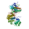
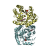
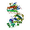
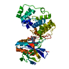
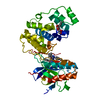
 Lactobacillus casei ATCC 334 (bacteria)
 X-RAY DIFFRACTION /
  Authors
 Citation
 Structure visualization
 Downloads & links
 Other downloads


 PDBj
PDBj

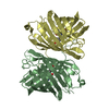
 Assembly
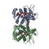

 Mass: 745.421 Da / Num. of mol.: 2 / Source method: obtained synthetically / Formula: C21H30N7O17P3
 Mass: 18.015 Da / Num. of mol.: 360 / Source method: isolated from a natural source / Formula: H2O
 Sample preparation
 / Beamline: X4A / Wavelength: 0.97853 Å
 Processing