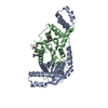
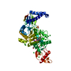
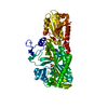
- PDB-3h0n: Crystal structure of a duf1470 family protein (jann_2411) from ja... -
+
Open data
ID or keywords:
Loading...
-
Basic information
Entry
Database: PDB / ID: 3h0n
Title
Crystal structure of a duf1470 family protein (jann_2411) from jannaschia sp. ccs1 at 1.45 A resolution
Components
uncharacterized protein DUF1470
Keywords
METAL BINDING PROTEIN / Treble clef zinc finger / structural genomics / Joint Center for Structural Genomics / JCSG / Protein Structure Initiative / PSI-2
Mass: 18.015 Da / Num. of mol.: 240 / Source method: isolated from a natural source / Formula: H2O
-
Details
Has protein modification
Y
Sequence details
THE CONSTRUCT WAS EXPRESSED WITH A PURIFICATION TAG MGSDKIHHHHHHENLYFQG. THE TAG WAS REMOVED WITH ...THE CONSTRUCT WAS EXPRESSED WITH A PURIFICATION TAG MGSDKIHHHHHHENLYFQG. THE TAG WAS REMOVED WITH TEV PROTEASE LEAVING ONLY A GLYCINE (0) FOLLOWED BY THE TARGET SEQUENCE.
-
Experimental details
-
Experiment
Experiment
Method: X-RAY DIFFRACTION / Number of used crystals: 1
-
Sample preparation
Crystal
Density Matthews: 2.51 Å3/Da / Density % sol: 50.95 %
Crystal grow
Temperature: 277 K / Method: vapor diffusion, sitting drop / pH: 6.5 Details: 1.40M sodium acetate, 0.10M sodium cacodylate pH 6.5, NANODROP, VAPOR DIFFUSION, SITTING DROP, temperature 277K
Monochromator: Single crystal Si(111) bent monochromator (horizontal focusing) Protocol: MAD / Monochromatic (M) / Laue (L): M / Scattering type: x-ray
Radiation wavelength
ID
Wavelength (Å)
Relative weight
1
0.91837
1
2
0.9792
1
3
0.97879
1
Reflection
Resolution: 1.45→25.786 Å / Num. obs: 36254 / % possible obs: 97.7 % / Observed criterion σ(I): -3 / Biso Wilson estimate: 13.251 Å2 / Rmerge(I) obs: 0.028 / Net I/σ(I): 14.38
Reflection shell
Resolution (Å)
Rmerge(I) obs
Mean I/σ(I) obs
Num. measured obs
Num. unique obs
Diffraction-ID
% possible all
1.45-1.5
0.249
2.8
6683
6767
1
97.1
1.5-1.56
0.193
3.8
7187
7165
1
99
1.56-1.63
0.14
4.7
7160
7076
1
99.2
1.63-1.72
0.114
6
7644
7430
1
98.9
1.72-1.83
0.085
8.1
7599
7268
1
98.8
1.83-1.97
0.057
11.2
7536
7040
1
98.4
1.97-2.17
0.035
16.8
7793
7091
1
98
2.17-2.48
0.026
21.8
7797
6951
1
97.7
2.48-3.12
0.02
29.1
8069
6937
1
96.5
3.12-25.786
0.013
40.9
8425
6862
1
93.8
-
Phasing
Phasing
Method: MAD
-
Processing
Software
Name
Version
Classification
NB
REFMAC
5.5.0053
refinement
PHENIX
refinement
SOLVE
phasing
MolProbity
3beta29
modelbuilding
XSCALE
datascaling
PDB_EXTRACT
3.006
dataextraction
XDS
datareduction
Refinement
Method to determine structure: MAD / Resolution: 1.45→25.786 Å / Cor.coef. Fo:Fc: 0.973 / Cor.coef. Fo:Fc free: 0.97 / Occupancy max: 1 / Occupancy min: 0.3 / SU B: 1.706 / SU ML: 0.033 / TLS residual ADP flag: LIKELY RESIDUAL / Cross valid method: THROUGHOUT / σ(F): 0 / ESU R: 0.053 / ESU R Free: 0.053 Stereochemistry target values: MAXIMUM LIKELIHOOD WITH PHASES Details: 1.HYDROGENS HAVE BEEN ADDED IN THE RIDING POSITIONS. 2.ATOM RECORD CONTAINS RESIDUAL B FACTORS ONLY. 3.A MET-INHIBITION PROTOCOL WAS USED FOR SELENOMETHIONINE INCORPORATION DURING PROTEIN ...Details: 1.HYDROGENS HAVE BEEN ADDED IN THE RIDING POSITIONS. 2.ATOM RECORD CONTAINS RESIDUAL B FACTORS ONLY. 3.A MET-INHIBITION PROTOCOL WAS USED FOR SELENOMETHIONINE INCORPORATION DURING PROTEIN EXPRESSION. THE OCCUPANCY OF THE SE ATOMS IN THE MSE RESIDUES WAS REDUCED TO 0.75 TO ACCOUNT FOR THE REDUCED SCATTERING POWER DUE TO PARTIAL S-MET INCORPORATION. 4.ONE ZN ION, ONE NI ION, TWO NA IONS, THREE ACETATE IONS AND TWO GLYCEROL MOLECULES WERE MODELED. THE PRESENCE OF THE ZN AND NI ATOMS ARE SUPPORTED BY X-RAY FLUORESCENCE MEASUREMENTS, ANOMALOUS DIFFERENCE FOURIERS ABOVE AND BELOW THE NI AND ZN ABSORPTION EDGES AND GEOMETRY. 5.RESIDUES 0 AND 185 TO 187 ARE DISORDERED AND NOT MODELED IN THE STRUCTURE.
Rfactor
Num. reflection
% reflection
Selection details
Rfree
0.157
1810
5 %
RANDOM
Rwork
0.14
-
-
-
obs
0.141
36254
99.14 %
-
Solvent computation
Ion probe radii: 0.8 Å / Shrinkage radii: 0.8 Å / VDW probe radii: 1.4 Å / Solvent model: MASK
In the structure databanks used in Yorodumi, some data are registered as the other names, "COVID-19 virus" and "2019-nCoV". Here are the details of the virus and the list of structure data.
Jan 31, 2019. EMDB accession codes are about to change! (news from PDBe EMDB page)
EMDB accession codes are about to change! (news from PDBe EMDB page)
The allocation of 4 digits for EMDB accession codes will soon come to an end. Whilst these codes will remain in use, new EMDB accession codes will include an additional digit and will expand incrementally as the available range of codes is exhausted. The current 4-digit format prefixed with “EMD-” (i.e. EMD-XXXX) will advance to a 5-digit format (i.e. EMD-XXXXX), and so on. It is currently estimated that the 4-digit codes will be depleted around Spring 2019, at which point the 5-digit format will come into force.
The EM Navigator/Yorodumi systems omit the EMD- prefix.
Related info.:Q: What is EMD? / ID/Accession-code notation in Yorodumi/EM Navigator
Yorodumi is a browser for structure data from EMDB, PDB, SASBDB, etc.
This page is also the successor to EM Navigator detail page, and also detail information page/front-end page for Omokage search.
The word "yorodu" (or yorozu) is an old Japanese word meaning "ten thousand". "mi" (miru) is to see.
Related info.:EMDB / PDB / SASBDB / Comparison of 3 databanks / Yorodumi Search / Aug 31, 2016. New EM Navigator & Yorodumi / Yorodumi Papers / Jmol/JSmol / Function and homology information / Changes in new EM Navigator and Yorodumi
 Movie
Movie Controller

 Yorodumi
 Open data
 Basic information
 Components
 Keywords
 Function and homology information
 Jannaschia sp. CCS1 (bacteria)
 X-RAY DIFFRACTION /
  Authors
 Citation
 Structure visualization
 Downloads & links
 Other downloads
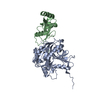


 PDBj
PDBj Assembly


 Mass: 65.409 Da / Num. of mol.: 1 / Source method: obtained synthetically / Formula: Zn
 Mass: 58.693 Da / Num. of mol.: 1 / Source method: obtained synthetically / Formula: Ni
 Mass: 22.990 Da / Num. of mol.: 2 / Source method: obtained synthetically / Formula: Na
 Mass: 92.094 Da / Num. of mol.: 2 / Source method: obtained synthetically / Formula: C3H8O3
 Mass: 59.044 Da / Num. of mol.: 3 / Source method: obtained synthetically / Formula: C2H3O2
 Sample preparation
 / Beamline: BL11-1 / Wavelength: 0.91837,0.97920,0.97879
 Processing