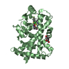
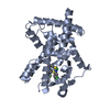
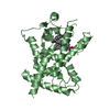
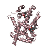
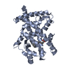
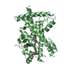
Entry Database : PDB / ID : 3gz9Title Crystal Structure of Peroxisome Proliferator-Activated Receptor Delta (PPARd) in Complex with a Full Agonist Peroxisome proliferator-activated receptor delta Keywords / / / / / / / / / / / / / / / Function / homology Function Domain/homology Component
/ / / / / / / / / / / / / / / / / / / / / / / / / / / / / / / / / / / / / / / / / / / / / / / / / / / / / / / / / / / / / / / / / / / / / / / / / / / / / / / / / / / / / / / / / / / / / / / / / / / / / / / / / / / / / / / / / / / / / Biological species Homo sapiens (human)Method / / / Resolution : 2 Å Authors Wang, Z. / Sudom, A. / Walker, N.P. Journal : Bioorg.Med.Chem.Lett. / Year : 2009Title : Identification of a PPARdelta agonist with partial agonistic activity on PPARgamma.Authors: Connors, R.V. / Wang, Z. / Harrison, M. / Zhang, A. / Wanska, M. / Hiscock, S. / Fox, B. / Dore, M. / Labelle, M. / Sudom, A. / Johnstone, S. / Liu, J. / Walker, N.P. / Chai, A. / Siegler, K. ... Authors : Connors, R.V. / Wang, Z. / Harrison, M. / Zhang, A. / Wanska, M. / Hiscock, S. / Fox, B. / Dore, M. / Labelle, M. / Sudom, A. / Johnstone, S. / Liu, J. / Walker, N.P. / Chai, A. / Siegler, K. / Li, Y. / Coward, P. History Deposition Apr 6, 2009 Deposition site / Processing site Revision 1.0 Jun 30, 2009 Provider / Type Revision 1.1 Jul 13, 2011 Group Revision 1.2 Jul 29, 2020 Group / Derived calculations / Structure summaryCategory chem_comp / entity ... chem_comp / entity / pdbx_chem_comp_identifier / pdbx_entity_nonpoly / struct_site / struct_site_gen Item _chem_comp.mon_nstd_flag / _chem_comp.name ... _chem_comp.mon_nstd_flag / _chem_comp.name / _chem_comp.type / _entity.pdbx_description / _pdbx_entity_nonpoly.name Description / Provider / Type Revision 1.3 Oct 13, 2021 Group / Structure summary / Category / database_2 / struct_ref_seq_difItem _chem_comp.pdbx_synonyms / _database_2.pdbx_DOI ... _chem_comp.pdbx_synonyms / _database_2.pdbx_DOI / _database_2.pdbx_database_accession / _struct_ref_seq_dif.details Revision 1.4 Feb 21, 2024 Group / Category / chem_comp_bond
Show all Show less
 Movie
Movie Controller

 Yorodumi
 Open data
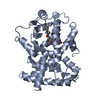
 Basic information
 Components
 Keywords
 Function and homology information
 Homo sapiens (human)
 X-RAY DIFFRACTION /
  Authors
 Citation
 Structure visualization
 Downloads & links
 Other downloads
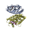

 PDBj
PDBj
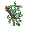
 Assembly

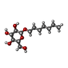
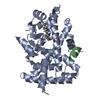
 Type: D-saccharide / Mass: 278.342 Da / Num. of mol.: 1 / Source method: obtained synthetically / Formula: C13H26O6
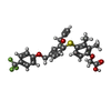
 Mass: 516.529 Da / Num. of mol.: 1 / Source method: obtained synthetically / Formula: C27H23F3O5S
 Mass: 18.015 Da / Num. of mol.: 172 / Source method: isolated from a natural source / Formula: H2O
 Sample preparation
 / Beamline: 5.0.2 / Wavelength: 1.541 Å
 Processing