 Movie
Movie Controller

[English] 日本語
 Yorodumi
- PDB-3gqm: Crystal structure of Cell Inhibiting Factor (Cif) from Burkholder... -
+ Open data

- Basic information
| Entry | Database: PDB / ID: 3gqm | ||||||
|---|---|---|---|---|---|---|---|
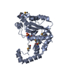
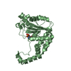
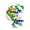
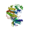
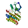
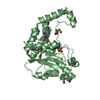
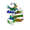
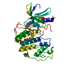
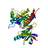
| Title | Crystal structure of Cell Inhibiting Factor (Cif) from Burkholderia pseudomallei (CifBp) | ||||||
 Components Components | Cell Inhibiting Factor (CifBp) | ||||||
 Keywords Keywords | UNKNOWN FUNCTION / Cell Inhibiting factor / Cif / CifBp | ||||||
| Function / homology |  Function and homology information Function and homology informationprotein-glutamine glutaminase activity / protein-glutamine glutaminase / symbiont-mediated perturbation of host cell cycle progression / toxin activity / symbiont-mediated suppression of host NF-kappaB cascade / host cell nucleus / extracellular region Similarity search - Function | ||||||
| Biological species |  Burkholderia pseudomallei (bacteria) Burkholderia pseudomallei (bacteria) | ||||||
| Method |  X-RAY DIFFRACTION / SYNCHROTRON / MOLECULAR REPLACEMENT / Resolution: 2.1 Å X-RAY DIFFRACTION / SYNCHROTRON / MOLECULAR REPLACEMENT / Resolution: 2.1 Å | ||||||
 Authors Authors | Crow, A. / Banfield, M.J. | ||||||
 Citation Citation | Journal: Plos One / Year: 2009 Title: Crystal structures of Cif from bacterial pathogens Photorhabdus luminescens and Burkholderia pseudomallei. Authors: Crow, A. / Race, P.R. / Jubelin, G. / Varela Chavez, C. / Escoubas, J.M. / Oswald, E. / Banfield, M.J. | ||||||
| History |
| ||||||
| Remark 650 | HELIX DETERMINATION METHOD: AUTHOR DETERMINED |
- Structure visualization
| Structure viewer | Molecule: MolmilJmol/JSmol |
|---|
- Downloads & links
-Download
| PDBx/mmCIF format | 3gqm.cif.gz | 110.1 KB | Display | PDBx/mmCIF format |
|---|---|---|---|---|
| PDB format | pdb3gqm.ent.gz | 84.1 KB | Display | PDB format |
| PDBx/mmJSON format | 3gqm.json.gz | Tree view | PDBx/mmJSON format | |
| Others |  Other downloads Other downloads |
-Validation report
| Arichive directory | https://data.pdbj.org/pub/pdb/validation_reports/gq/3gqmftp://data.pdbj.org/pub/pdb/validation_reports/gq/3gqm | HTTPS FTP |
|---|
-Related structure data
| Related structure data | 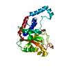 3gqjSC S: Starting model for refinement C: citing same article ( |
|---|---|
| Similar structure data |


-Links
 PDBj
PDBj
- Assembly
| Deposited unit | 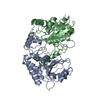
| ||||||||
|---|---|---|---|---|---|---|---|---|---|
| 1 | 
| ||||||||
| 2 | 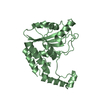
| ||||||||
| Unit cell |
|
-Components
| #1: Protein | Mass: 30854.035 Da / Num. of mol.: 2 Source method: isolated from a genetically manipulated source Source: (gene. exp.) Burkholderia pseudomallei (bacteria) / Gene: BPSS1385 / Production host: #2: Water | ChemComp-HOH / |  Mass: 18.015 Da / Num. of mol.: 92 / Source method: isolated from a natural source / Formula: H2O Mass: 18.015 Da / Num. of mol.: 92 / Source method: isolated from a natural source / Formula: H2O |
|---|
-Experimental details
-Experiment
| Experiment | Method: X-RAY DIFFRACTION / Number of used crystals: 1 |
|---|
- Sample preparation
| Crystal | Density Matthews: 1.89 Å3/Da / Density % sol: 35.07 % |
|---|---|
| Crystal grow | Temperature: 291 K / Method: vapor diffusion, sitting drop / pH: 7.5 Details: 10 % (w/v) PEG 8000, 100 mM HEPES 7.5, VAPOR DIFFUSION, SITTING DROP, temperature 291K |
-Data collection
| Diffraction | Mean temperature: 100 K |
|---|---|
| Diffraction source | Source: SYNCHROTRON / Site: SRS  / Beamline: PX9.6 / Wavelength: 0.92 Å / Beamline: PX9.6 / Wavelength: 0.92 Å |
| Detector | Detector: CCD / Date: Jun 6, 2008 |
| Radiation | Protocol: SINGLE WAVELENGTH / Monochromatic (M) / Laue (L): M / Scattering type: x-ray |
| Radiation wavelength | Wavelength: 0.92 Å / Relative weight: 1 |
| Reflection | Resolution: 2.1→22.01 Å / Num. all: 29440 / Num. obs: 29440 / % possible obs: 99.9 % / Observed criterion σ(F): 1 / Observed criterion σ(I): 1 / Redundancy: 5 % / Rsym value: 0.05 / Net I/σ(I): 20.2 |
| Reflection shell | Resolution: 2.1→2.21 Å / Redundancy: 4.9 % / Mean I/σ(I) obs: 3.5 / Rsym value: 0.374 / % possible all: 99.9 |
- Processing
| Software |
| ||||||||||||||||||||||||||||||||||||||||||||||||||||||||||||||||||||||||||||||||||||||||||||||||||||||||||||||||||||||||||||||||||||||||||||||||||||||||||||||||||||||||||
|---|---|---|---|---|---|---|---|---|---|---|---|---|---|---|---|---|---|---|---|---|---|---|---|---|---|---|---|---|---|---|---|---|---|---|---|---|---|---|---|---|---|---|---|---|---|---|---|---|---|---|---|---|---|---|---|---|---|---|---|---|---|---|---|---|---|---|---|---|---|---|---|---|---|---|---|---|---|---|---|---|---|---|---|---|---|---|---|---|---|---|---|---|---|---|---|---|---|---|---|---|---|---|---|---|---|---|---|---|---|---|---|---|---|---|---|---|---|---|---|---|---|---|---|---|---|---|---|---|---|---|---|---|---|---|---|---|---|---|---|---|---|---|---|---|---|---|---|---|---|---|---|---|---|---|---|---|---|---|---|---|---|---|---|---|---|---|---|---|---|---|---|
| Refinement | Method to determine structure: MOLECULAR REPLACEMENT Starting model: 3GQJ Resolution: 2.1→22.01 Å / Cor.coef. Fo:Fc: 0.943 / Cor.coef. Fo:Fc free: 0.911 / Cross valid method: THROUGHOUT / σ(F): 0 / ESU R: 0.256 / ESU R Free: 0.208 / Stereochemistry target values: MAXIMUM LIKELIHOOD / Details: HYDROGENS HAVE BEEN ADDED IN THE RIDING POSITIONS
| ||||||||||||||||||||||||||||||||||||||||||||||||||||||||||||||||||||||||||||||||||||||||||||||||||||||||||||||||||||||||||||||||||||||||||||||||||||||||||||||||||||||||||
| Solvent computation | Ion probe radii: 0.8 Å / Shrinkage radii: 0.8 Å / VDW probe radii: 1.2 Å / Solvent model: MASK | ||||||||||||||||||||||||||||||||||||||||||||||||||||||||||||||||||||||||||||||||||||||||||||||||||||||||||||||||||||||||||||||||||||||||||||||||||||||||||||||||||||||||||
| Displacement parameters | Biso mean: 36.052 Å2
| ||||||||||||||||||||||||||||||||||||||||||||||||||||||||||||||||||||||||||||||||||||||||||||||||||||||||||||||||||||||||||||||||||||||||||||||||||||||||||||||||||||||||||
| Refinement step | Cycle: LAST / Resolution: 2.1→22.01 Å
| ||||||||||||||||||||||||||||||||||||||||||||||||||||||||||||||||||||||||||||||||||||||||||||||||||||||||||||||||||||||||||||||||||||||||||||||||||||||||||||||||||||||||||
| Refine LS restraints |
| ||||||||||||||||||||||||||||||||||||||||||||||||||||||||||||||||||||||||||||||||||||||||||||||||||||||||||||||||||||||||||||||||||||||||||||||||||||||||||||||||||||||||||
| LS refinement shell | Resolution: 2.1→2.155 Å / Total num. of bins used: 20
|
