 Movie
Movie Controller

[English] 日本語
 Yorodumi
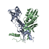
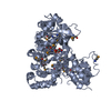
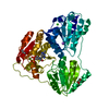
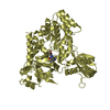
- PDB-3gpl: Crystal structure of the ternary complex of RecD2 with DNA and ADPNP -
+ Open data

- Basic information
| Entry | Database: PDB / ID: 3gpl | ||||||
|---|---|---|---|---|---|---|---|
| Title | Crystal structure of the ternary complex of RecD2 with DNA and ADPNP | ||||||
 Components Components |
| ||||||
 Keywords Keywords | HYDROLASE/DNA / ALPHA AND BETA PROTEIN / ATP-BINDING / NUCLEOTIDE-BINDING / HELICASE / HYDROLASE-DNA COMPLEX | ||||||
| Function / homology |  Function and homology information Function and homology informationexodeoxyribonuclease V complex / DNA 5'-3' helicase / single-stranded DNA helicase activity / ATP-dependent activity, acting on DNA / 5'-3' DNA helicase activity / DNA recombination / ATP hydrolysis activity / DNA binding / ATP binding Similarity search - Function | ||||||
| Biological species |  Deinococcus radiodurans R1 (radioresistant) Deinococcus radiodurans R1 (radioresistant) | ||||||
| Method |  X-RAY DIFFRACTION / MOLECULAR REPLACEMENT / Resolution: 2.5 Å X-RAY DIFFRACTION / MOLECULAR REPLACEMENT / Resolution: 2.5 Å | ||||||
 Authors Authors | Saikrishnan, K. / Cook, N. / Wigley, D.B. | ||||||
 Citation Citation | Journal: Cell(Cambridge,Mass.) / Year: 2009 Title: Mechanistic basis of 5'-3' translocation in SF1B helicases. Authors: Saikrishnan, K. / Powell, B. / Cook, N.J. / Webb, M.R. / Wigley, D.B. #1: Journal: Embo J. / Year: 2008Title: DNA binding to RecD: role of the 1B domain in SF1B helicase activity. Authors: Saikrishnan, K. / Griffiths, S.P. / Cook, N. / Court, R. / Wigley, D.B. | ||||||
| History |
|
- Structure visualization
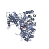
| Structure viewer | Molecule: MolmilJmol/JSmol |
|---|
- Downloads & links
-Download
| PDBx/mmCIF format | 3gpl.cif.gz | 220.3 KB | Display | PDBx/mmCIF format |
|---|---|---|---|---|
| PDB format | pdb3gpl.ent.gz | 173.1 KB | Display | PDB format |
| PDBx/mmJSON format | 3gpl.json.gz | Tree view | PDBx/mmJSON format | |
| Others |  Other downloads Other downloads |
-Validation report
| Summary document | 3gpl_validation.pdf.gz | 1.1 MB | Display | wwPDB validaton report |
|---|---|---|---|---|
| Full document | 3gpl_full_validation.pdf.gz | 1.1 MB | Display | |
| Data in XML | 3gpl_validation.xml.gz | 43.7 KB | Display | |
| Data in CIF | 3gpl_validation.cif.gz | 60.2 KB | Display | |
| Arichive directory | https://data.pdbj.org/pub/pdb/validation_reports/gp/3gplftp://data.pdbj.org/pub/pdb/validation_reports/gp/3gpl | HTTPS FTP |
-Related structure data
| Related structure data |  3gp8C  3e1sS C: citing same article ( S: Starting model for refinement |
|---|---|
| Similar structure data |


-Links
 PDBj
PDBj


- Assembly
| Deposited unit | 
| ||||||||
|---|---|---|---|---|---|---|---|---|---|
| 1 | 
| ||||||||
| 2 | 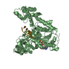
| ||||||||
| Unit cell |
|
-Components
| #1: Protein | Mass: 61465.914 Da / Num. of mol.: 2 / Fragment: UNP residues 151-715 Source method: isolated from a genetically manipulated source Details: N-terminus deletion mutant of RecD2 Source: (gene. exp.) Deinococcus radiodurans R1 (radioresistant)Strain: R1 / DSM 20539 / IFO 15346 / LMG 4051 / NCIB 9279 / Gene: DR_1902, recD2 / Plasmid: pET22b / Production host: #2: DNA chain | Mass: 2388.585 Da / Num. of mol.: 2 / Source method: obtained synthetically / Details: Synthetic DNA #3: Chemical |   Mass: 24.305 Da / Num. of mol.: 2 / Source method: obtained synthetically / Formula: Mg Mass: 24.305 Da / Num. of mol.: 2 / Source method: obtained synthetically / Formula: Mg#4: Chemical |   Mass: 506.196 Da / Num. of mol.: 2 / Source method: obtained synthetically / Formula: C10H17N6O12P3 / Comment: AMP-PNP, energy-carrying molecule analogue*YM Mass: 506.196 Da / Num. of mol.: 2 / Source method: obtained synthetically / Formula: C10H17N6O12P3 / Comment: AMP-PNP, energy-carrying molecule analogue*YM#5: Water | ChemComp-HOH / |  Mass: 18.015 Da / Num. of mol.: 186 / Source method: isolated from a natural source / Formula: H2O Mass: 18.015 Da / Num. of mol.: 186 / Source method: isolated from a natural source / Formula: H2O |
|---|
-Experimental details
-Experiment
| Experiment | Method: X-RAY DIFFRACTION / Number of used crystals: 1 |
|---|
- Sample preparation
| Crystal | Density Matthews: 2.58 Å3/Da / Density % sol: 52.3 % | ||||||||||||||||||||
|---|---|---|---|---|---|---|---|---|---|---|---|---|---|---|---|---|---|---|---|---|---|
| Crystal grow | Temperature: 298 K / Method: vapor diffusion, hanging drop / pH: 8.5 Details: 10% PEG 8000, 100 mM Tris-HCl pH 8.5, VAPOR DIFFUSION, HANGING DROP, temperature 298K | ||||||||||||||||||||
| Components of the solutions |
|
-Data collection
| Diffraction | Mean temperature: 100 K |
|---|---|
| Diffraction source | Source: ROTATING ANODE / Type: RIGAKU MICROMAX-007 / Wavelength: 1.5418 Å |
| Detector | Type: MAR scanner 345 mm plate / Detector: IMAGE PLATE / Date: Jun 20, 2008 / Details: mirrors |
| Radiation | Monochromator: Varimax / Protocol: SINGLE WAVELENGTH / Monochromatic (M) / Laue (L): M / Scattering type: x-ray |
| Radiation wavelength | Wavelength: 1.5418 Å / Relative weight: 1 |
| Reflection | Resolution: 2.5→50 Å / Num. all: 44807 / Num. obs: 44090 / % possible obs: 98.4 % / Observed criterion σ(I): -3 / Redundancy: 2.3 % / Biso Wilson estimate: 27.2 Å2 / Rmerge(I) obs: 0.047 / Rsym value: 0.047 / Net I/σ(I): 16.05 |
| Reflection shell | Resolution: 2.5→2.6 Å / Redundancy: 2 % / Rmerge(I) obs: 0.251 / Mean I/σ(I) obs: 3.83 / Num. unique all: 4633 / Rsym value: 0.251 / % possible all: 94.6 |
- Processing
| Software |
| ||||||||||||||||||||||||||||||||||||||||||||||||||||||||||||
|---|---|---|---|---|---|---|---|---|---|---|---|---|---|---|---|---|---|---|---|---|---|---|---|---|---|---|---|---|---|---|---|---|---|---|---|---|---|---|---|---|---|---|---|---|---|---|---|---|---|---|---|---|---|---|---|---|---|---|---|---|---|
| Refinement | Method to determine structure: MOLECULAR REPLACEMENT Starting model: PDB entry 3E1S Resolution: 2.5→43.27 Å / Rfactor Rfree error: 0.006 / Data cutoff high absF: 1445846.88 / Data cutoff low absF: 0 / Isotropic thermal model: RESTRAINED / Cross valid method: THROUGHOUT / σ(F): 0 / Stereochemistry target values: Engh & Huber
| ||||||||||||||||||||||||||||||||||||||||||||||||||||||||||||
| Solvent computation | Solvent model: FLAT MODEL / Bsol: 33.1733 Å2 / ksol: 0.35 e/Å3 | ||||||||||||||||||||||||||||||||||||||||||||||||||||||||||||
| Displacement parameters | Biso mean: 45.6 Å2
| ||||||||||||||||||||||||||||||||||||||||||||||||||||||||||||
| Refine analyze |
| ||||||||||||||||||||||||||||||||||||||||||||||||||||||||||||
| Refinement step | Cycle: LAST / Resolution: 2.5→43.27 Å
| ||||||||||||||||||||||||||||||||||||||||||||||||||||||||||||
| Refine LS restraints |
| ||||||||||||||||||||||||||||||||||||||||||||||||||||||||||||
| LS refinement shell | Resolution: 2.5→2.66 Å / Rfactor Rfree error: 0.019 / Total num. of bins used: 6
|
