Mass: 18.015 Da / Num. of mol.: 192 / Source method: isolated from a natural source / Formula: H2O
-
Details
Has protein modification
Y
Sequence details
THE CONSTRUCT WAS EXPRESSED WITH A PURIFICATION TAG MGSDKIHHHHHHENLYFQG. THE TAG WAS REMOVED WITH ...THE CONSTRUCT WAS EXPRESSED WITH A PURIFICATION TAG MGSDKIHHHHHHENLYFQG. THE TAG WAS REMOVED WITH TEV PROTEASE LEAVING ONLY A GLYCINE (0) FOLLOWED BY THE TARGET SEQUENCE.
-
Experimental details
-
Experiment
Experiment
Method: X-RAY DIFFRACTION / Number of used crystals: 1
-
Sample preparation
Crystal
Description: DATA WERE SCALED USING XSCALE WITH FRIEDEL PAIRS KEPT AS SEPARATE WHEN COMPUTING R MERGE, COMPLETENESS AND .
Crystal grow
Temperature: 293 K / Method: vapor diffusion, sitting drop / pH: 6.5 Details: 0.01M cobalt chloride, 1.8M ammonium sulfate, 0.1M MES pH 6.5, ADDITIVE: 0.001 M S-ADENOSYLMETHIONINE (SAM), NANODROP', VAPOR DIFFUSION, SITTING DROP, temperature 293K
Resolution: 2.11→30.083 Å / Num. obs: 42096 / % possible obs: 99.7 % / Observed criterion σ(I): -3 / Redundancy: 17.9 % / Biso Wilson estimate: 40.16 Å2 / Rmerge(I) obs: 0.221 / Net I/σ(I): 11
Reflection shell
Resolution (Å)
Rmerge(I) obs
Mean I/σ(I) obs
Num. measured obs
Num. unique obs
Diffraction-ID
% possible all
2.11-2.19
1.53
2
64498
8394
1
99.6
2.19-2.27
1.15
2.7
54873
7150
1
99.4
2.27-2.38
0.91
3.5
64861
8442
1
99.7
2.38-2.5
0.67
4.9
58989
7574
1
99.8
2.5-2.66
0.81
6.3
72014
8105
1
99.8
2.66-2.86
0.65
8.4
75514
7751
1
99.8
2.86-3.15
0.53
12.2
87968
7989
1
99.6
3.15-3.6
0.32
17.8
88970
7903
1
99.9
3.6-4.53
0.15
24.2
90866
7956
1
99.8
4.53-30.083
0.09
27.8
93659
8021
1
99.5
-
Phasing
Phasing
Method: MAD
-
Processing
Software
Name
Version
Classification
NB
REFMAC
5.2.0019
refinement
PHENIX
refinement
SHELX
phasing
MolProbity
3beta29
modelbuilding
XSCALE
datascaling
PDB_EXTRACT
3.006
dataextraction
XDS
datareduction
SHELXD
phasing
autoSHARP
phasing
Refinement
Method to determine structure: MAD / Resolution: 2.11→30.083 Å / Cor.coef. Fo:Fc: 0.969 / Cor.coef. Fo:Fc free: 0.967 / Occupancy max: 1 / Occupancy min: 0.5 / SU B: 4.175 / SU ML: 0.062 / TLS residual ADP flag: LIKELY RESIDUAL / Cross valid method: THROUGHOUT / σ(F): 0 / ESU R: 0.095 / ESU R Free: 0.09 Stereochemistry target values: MAXIMUM LIKELIHOOD WITH PHASES Details: 1.HYDROGENS HAVE BEEN ADDED IN THE RIDING POSITIONS. 2.ATOM RECORD CONTAINS RESIDUAL B FACTORS ONLY. 3.A MET-INHIBITION PROTOCOL WAS USED FOR SELENOMETHIONINE INCORPORATION DURING PROTEIN ...Details: 1.HYDROGENS HAVE BEEN ADDED IN THE RIDING POSITIONS. 2.ATOM RECORD CONTAINS RESIDUAL B FACTORS ONLY. 3.A MET-INHIBITION PROTOCOL WAS USED FOR SELENOMETHIONINE INCORPORATION DURING PROTEIN EXPRESSION. THE OCCUPANCY OF THE SE ATOMS IN THE MSE RESIDUES WAS REDUCED TO 0.75 TO ACCOUNT FOR THE REDUCED SCATTERING POWER DUE TO PARTIAL S-MET INCORPORATION. 4.S-ADENOSYLMETHIONINE WAS ADDED DURING CRYSTALLIZATION. IN THIS STRUCTURE, ONLY S-ADENOSYL-L-HOMOCYSTEINE( SAH) IS MODELED IN THE CONSERVED BINDING SITE DUE TO A POSSIBLE ENZYMATICALLY RELATED DEGRADATION. AN UNKNOWN LIGAND (UNL) IS MODELED NEXT TO THE S-ADENOSYL-L- HOMOCYSTEINE IN THE SAME BINDING SITE. THE SHAPE OF THE LIGAND IS CREATINE LIKE. 5.A COBALT ION FROM THE CRYSTALLIZATION REAGENT IS MODELED IN THIS STRUCTURE. THE PRESENCE OF THE CO ATOM IS SUPPORTED BY X-RAY FLUORESCENCE MEASUREMENTS, ANOMALOUS DIFFERENCE FOURIERS AND GEOMETRY. ADDITIONAL ELECTRON DENSITY IS OBSERVED ADJACENT TO THE COBALT ION AND NEAR TO RESIDUES 203 AND 243. THIS DENSITY WAS LEFT UNMODELED. 6.AN IMIDAZOLE MOLECULE AND CHLORIDE ION FROM THE PROTEIN BUFFER, SULFATE IONS FROM THE CRYSTALLIZATION CONDITION AND ETHYLENE GLYCOL MOLECULES FROM THE CRYOPROTECTANT ARE ALSO MODELED IN THIS STRUCTURE.
Rfactor
Num. reflection
% reflection
Selection details
Rfree
0.175
2128
5.1 %
RANDOM
Rwork
0.162
-
-
-
obs
0.163
42037
99.77 %
-
Solvent computation
Ion probe radii: 0.8 Å / Shrinkage radii: 0.8 Å / VDW probe radii: 1.2 Å / Solvent model: BABINET MODEL WITH MASK
In the structure databanks used in Yorodumi, some data are registered as the other names, "COVID-19 virus" and "2019-nCoV". Here are the details of the virus and the list of structure data.
Jan 31, 2019. EMDB accession codes are about to change! (news from PDBe EMDB page)
EMDB accession codes are about to change! (news from PDBe EMDB page)
The allocation of 4 digits for EMDB accession codes will soon come to an end. Whilst these codes will remain in use, new EMDB accession codes will include an additional digit and will expand incrementally as the available range of codes is exhausted. The current 4-digit format prefixed with “EMD-” (i.e. EMD-XXXX) will advance to a 5-digit format (i.e. EMD-XXXXX), and so on. It is currently estimated that the 4-digit codes will be depleted around Spring 2019, at which point the 5-digit format will come into force.
The EM Navigator/Yorodumi systems omit the EMD- prefix.
Related info.:Q: What is EMD? / ID/Accession-code notation in Yorodumi/EM Navigator
Yorodumi is a browser for structure data from EMDB, PDB, SASBDB, etc.
This page is also the successor to EM Navigator detail page, and also detail information page/front-end page for Omokage search.
The word "yorodu" (or yorozu) is an old Japanese word meaning "ten thousand". "mi" (miru) is to see.
Related info.:EMDB / PDB / SASBDB / Comparison of 3 databanks / Yorodumi Search / Aug 31, 2016. New EM Navigator & Yorodumi / Yorodumi Papers / Jmol/JSmol / Function and homology information / Changes in new EM Navigator and Yorodumi
 Movie
Movie Controller

 Yorodumi
 Open data
 Basic information
 Components
 Keywords
 Function and homology information
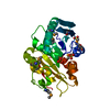
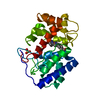
 Anabaena variabilis ATCC 29413 (bacteria)
 X-RAY DIFFRACTION /
  Authors
 Citation
 Structure visualization
 Downloads & links
 Other downloads


 PDBj
PDBj


 Assembly


 Mass: 58.933 Da / Num. of mol.: 1 / Source method: obtained synthetically / Formula: Co
 Mass: 35.453 Da / Num. of mol.: 1 / Source method: obtained synthetically / Formula: Cl
 Mass: 384.411 Da / Num. of mol.: 1 / Source method: obtained synthetically / Formula: C14H20N6O5S
 Mass: 69.085 Da / Num. of mol.: 1 / Source method: obtained synthetically / Formula: C3H5N2
 Mass: 96.063 Da / Num. of mol.: 4 / Source method: obtained synthetically / Formula: SO4
 Mass: 62.068 Da / Num. of mol.: 9 / Source method: obtained synthetically / Formula: C2H6O2
 Sample preparation
 / Beamline: 23-ID-B / Wavelength: 0.94645,0.97967
 Processing