Mass: 18.015 Da / Num. of mol.: 489 / Source method: isolated from a natural source / Formula: H2O
-
Details
Has protein modification
Y
Sequence details
THE CONSTRUCT WAS EXPRESSED WITH A PURIFICATION TAG MGSDKIHHHHHHENLYFQG.
-
Experimental details
-
Experiment
Experiment
Method: X-RAY DIFFRACTION / Number of used crystals: 1
-
Sample preparation
Crystal
Density Matthews: 2.31 Å3/Da / Density % sol: 46.85 % Description: THE AND R MERGE VALUES REPORTED HERE ARE BASED ON TREATING FRIEDEL PAIRS AS SEPARATE REFLECTIONS.
Crystal grow
Temperature: 277 K / Method: vapor diffusion, sitting drop / pH: 8.5 Details: 2.0000M (NH4)2SO4, 0.1M TRIS pH 8.5, NANODROP, VAPOR DIFFUSION, SITTING DROP, temperature 277K
Type: MARMOSAIC 325 mm CCD / Detector: CCD / Date: Nov 14, 2008 / Details: Flat mirror (vertical focusing)
Radiation
Monochromator: Single crystal Si(111) bent monochromator (horizontal focusing) Protocol: MAD / Monochromatic (M) / Laue (L): M / Scattering type: x-ray
Radiation wavelength
ID
Wavelength (Å)
Relative weight
1
0.91837
1
2
0.9787
1
3
0.97833
1
Reflection
Resolution: 1.7→29.161 Å / Num. obs: 48232 / % possible obs: 99.5 % / Observed criterion σ(I): -3 / Biso Wilson estimate: 20.057 Å2 / Rmerge(I) obs: 0.066 / Net I/σ(I): 14.75
Reflection shell
Resolution (Å)
Rmerge(I) obs
Mean I/σ(I) obs
Num. measured obs
Num. unique obs
Diffraction-ID
% possible all
1.7-1.76
0.735
1.8
31307
8862
1
98
1.76-1.83
0.55
2.5
32871
9056
1
99.7
1.83-1.91
0.382
3.6
32542
8795
1
99.5
1.91-2.02
0.256
5.4
37255
9923
1
99.7
2.02-2.14
0.178
7.7
33123
8665
1
99.7
2.14-2.31
0.127
10.4
35723
9329
1
99.7
2.31-2.54
0.087
14.8
34889
9054
1
99.7
2.54-2.9
0.062
19.8
34555
8950
1
99.8
2.9-3.66
0.032
33.1
35982
9278
1
99.8
3.66-29.161
0.022
47.7
35536
9139
1
99.4
-
Phasing
Phasing
Method: MAD
-
Processing
Software
Name
Version
Classification
NB
REFMAC
5.2.0019
refinement
PHENIX
refinement
SHELX
phasing
MolProbity
3beta29
modelbuilding
XSCALE
datascaling
PDB_EXTRACT
3.006
dataextraction
XDS
datareduction
SHELXD
phasing
autoSHARP
phasing
Refinement
Method to determine structure: MAD / Resolution: 1.7→29.161 Å / Cor.coef. Fo:Fc: 0.971 / Cor.coef. Fo:Fc free: 0.963 / Occupancy max: 1 / Occupancy min: 0.5 / SU B: 2.841 / SU ML: 0.05 / TLS residual ADP flag: LIKELY RESIDUAL / Cross valid method: THROUGHOUT / σ(F): 0 / ESU R: 0.083 / ESU R Free: 0.082 Stereochemistry target values: MAXIMUM LIKELIHOOD WITH PHASES Details: 1. HYDROGENS HAVE BEEN ADDED IN THE RIDING POSITIONS. 2. ATOM RECORDS CONTAIN RESIDUAL B FACTORS ONLY. 3. A MET-INHIBITION PROTOCOL WAS USED FOR SELENOMETHIONINE INCORPORATION DURING PROTEIN ...Details: 1. HYDROGENS HAVE BEEN ADDED IN THE RIDING POSITIONS. 2. ATOM RECORDS CONTAIN RESIDUAL B FACTORS ONLY. 3. A MET-INHIBITION PROTOCOL WAS USED FOR SELENOMETHIONINE INCORPORATION DURING PROTEIN EXPRESSION. THE OCCUPANCY OF THE SE ATOMS IN THE MSE RESIDUES WAS REDUCED TO 0.80 FOR THE REDUCED SCATTERING POWER DUE TO PARTIAL S-MET INCORPORATION. 4. A FLAVIN MONONUCLEOTIDE (FMN) MOLECULE ARE MODELED INTO THE PUTATIVE ACTIVE SITE ON EACH MONOMER. DURING THE REFINEMENT, THE FMN RESTRAINTS DICTIONARY WAS MODIFIED TO ALLOW BENDING OF THE ISOALLOXAZINE RING ALONG THE N5-N10 VIRTUAL AXIS RESULTING IN AN IMPROVED FIT BETWEEN THE FNN COORDINATES AND ELECTRON DENSITY. 5. AN UNKNOWN LIGAND (UNL) MOLECULE IS MODELED INTO THE PUTATIVE ACTIVE SITE OF EACH MONOMER. EACH UNL IS LOCATED ABOVE THE ISOALLOXAZINE MOIETY OF EACH FMN AND ITS SHAPE IS SIMILAR TO EITHER BENZOATE/NITOBENZENE OR BENZOATE/NITROBENZENE DERIVATIVES. 6. SULFATE (SO4) ION AND ETHYLENE GLYCOL (EDO) ARE MODELED BASED ON CRYSTALLIZATION AND CRYO CONDITION. 7. THERE ARE UNEXPECTED DENSITIES FOUND NEAR G171 AND THESE WERE NOT MODELED.
Rfactor
Num. reflection
% reflection
Selection details
Rfree
0.172
2445
5.1 %
RANDOM
Rwork
0.145
-
-
-
obs
0.146
48177
99.72 %
-
Solvent computation
Ion probe radii: 0.8 Å / Shrinkage radii: 0.8 Å / VDW probe radii: 1.2 Å / Solvent model: MASK
In the structure databanks used in Yorodumi, some data are registered as the other names, "COVID-19 virus" and "2019-nCoV". Here are the details of the virus and the list of structure data.
Jan 31, 2019. EMDB accession codes are about to change! (news from PDBe EMDB page)
EMDB accession codes are about to change! (news from PDBe EMDB page)
The allocation of 4 digits for EMDB accession codes will soon come to an end. Whilst these codes will remain in use, new EMDB accession codes will include an additional digit and will expand incrementally as the available range of codes is exhausted. The current 4-digit format prefixed with “EMD-” (i.e. EMD-XXXX) will advance to a 5-digit format (i.e. EMD-XXXXX), and so on. It is currently estimated that the 4-digit codes will be depleted around Spring 2019, at which point the 5-digit format will come into force.
The EM Navigator/Yorodumi systems omit the EMD- prefix.
Related info.:Q: What is EMD? / ID/Accession-code notation in Yorodumi/EM Navigator
Yorodumi is a browser for structure data from EMDB, PDB, SASBDB, etc.
This page is also the successor to EM Navigator detail page, and also detail information page/front-end page for Omokage search.
The word "yorodu" (or yorozu) is an old Japanese word meaning "ten thousand". "mi" (miru) is to see.
Related info.:EMDB / PDB / SASBDB / Comparison of 3 databanks / Yorodumi Search / Aug 31, 2016. New EM Navigator & Yorodumi / Yorodumi Papers / Jmol/JSmol / Function and homology information / Changes in new EM Navigator and Yorodumi
 Movie
Movie Controller

 Yorodumi
 Open data
 Basic information
 Components
 Keywords
 Function and homology information
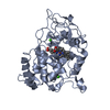
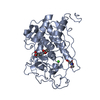
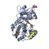
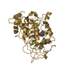
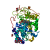
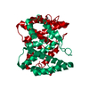
 Porphyromonas gingivalis W83 (bacteria)
 X-RAY DIFFRACTION /
  Authors
 Citation
 Structure visualization
 Downloads & links
 Other downloads


 PDBj
PDBj

 Assembly
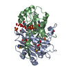


 Mass: 456.344 Da / Num. of mol.: 2 / Source method: obtained synthetically / Formula: C17H21N4O9P
 Mass: 96.063 Da / Num. of mol.: 3 / Source method: obtained synthetically / Formula: SO4
 Mass: 62.068 Da / Num. of mol.: 5 / Source method: obtained synthetically / Formula: C2H6O2
 Sample preparation
 / Beamline: BL11-1 / Wavelength: 0.91837,0.97870,0.97833
 Processing