 Movie
Movie Controller


+ Open data

- Basic information
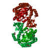
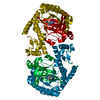
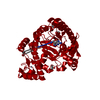
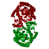
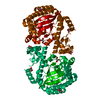
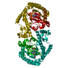
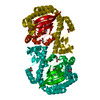
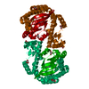
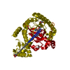
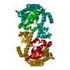
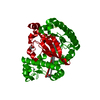
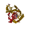
| Entry | Database: PDB / ID: 3gc4 | ||||||
|---|---|---|---|---|---|---|---|
| Title | tRNA-guanine transglycosylase in complex with inhibitor | ||||||
 Components Components | Queuine tRNA-ribosyltransferase | ||||||
 Keywords Keywords | TRANSFERASE / drug design / TIM Barrel / tRNA modification / Glycosyltransferase / Metal-binding / Queuosine biosynthesis / tRNA processing | ||||||
| Function / homology |  Function and homology information Function and homology informationtRNA-guanosine34 preQ1 transglycosylase / tRNA-guanosine(34) queuine transglycosylase activity / tRNA queuosine(34) biosynthetic process / metal ion binding / cytosol Similarity search - Function | ||||||
| Biological species |  Zymomonas mobilis (bacteria) Zymomonas mobilis (bacteria) | ||||||
| Method |  X-RAY DIFFRACTION / AB INITIO / Resolution: 1.8 Å X-RAY DIFFRACTION / AB INITIO / Resolution: 1.8 Å | ||||||
 Authors Authors | Ritschel, T. / Heine, A. / Klebe, G. | ||||||
 Citation Citation | Journal: Chemmedchem / Year: 2009 Title: How to Replace the Residual Solvation Shell of Polar Active Site Residues to Achieve Nanomolar Inhibition of tRNA-Guanine Transglycosylase Authors: Ritschel, T. / Kohler, P.C. / Neudert, G. / Heine, A. / Diederich, F. / Klebe, G. | ||||||
| History |
|
- Structure visualization
| Structure viewer | Molecule: MolmilJmol/JSmol |
|---|
- Downloads & links
-Download
| PDBx/mmCIF format | 3gc4.cif.gz | 90.8 KB | Display | PDBx/mmCIF format |
|---|---|---|---|---|
| PDB format | pdb3gc4.ent.gz | 65.8 KB | Display | PDB format |
| PDBx/mmJSON format | 3gc4.json.gz | Tree view | PDBx/mmJSON format | |
| Others |  Other downloads Other downloads |
-Validation report
| Arichive directory | https://data.pdbj.org/pub/pdb/validation_reports/gc/3gc4ftp://data.pdbj.org/pub/pdb/validation_reports/gc/3gc4 | HTTPS FTP |
|---|
-Related structure data
| Related structure data |  3eosC 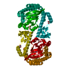 3eouC  3gc5C  3ge7C 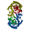 1p0eS C: citing same article ( S: Starting model for refinement |
|---|---|
| Similar structure data |


-Links
 PDBj
PDBj- Assembly
| Deposited unit | 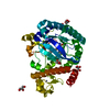
| ||||||||
|---|---|---|---|---|---|---|---|---|---|
| 1 | 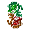
| ||||||||
| Unit cell |
|
-Components
| #1: Protein | Mass: 42925.703 Da / Num. of mol.: 1 Source method: isolated from a genetically manipulated source Source: (gene. exp.) Zymomonas mobilis (bacteria) / Gene: tgt, ZMO0363 / Plasmid: pET9d / Production host: References: UniProt: P28720, tRNA-guanosine34 preQ1 transglycosylase | ||||
|---|---|---|---|---|---|
| #2: Chemical | ChemComp-AAQ / 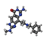  Mass: 363.416 Da / Num. of mol.: 1 / Source method: obtained synthetically / Formula: C19H21N7O Mass: 363.416 Da / Num. of mol.: 1 / Source method: obtained synthetically / Formula: C19H21N7O | ||||
| #3: Chemical | ChemComp-ZN /   Mass: 65.409 Da / Num. of mol.: 1 / Source method: obtained synthetically / Formula: Zn Mass: 65.409 Da / Num. of mol.: 1 / Source method: obtained synthetically / Formula: Zn | ||||
| #4: Chemical | ChemComp-GOL /   Mass: 92.094 Da / Num. of mol.: 5 / Source method: obtained synthetically / Formula: C3H8O3 Mass: 92.094 Da / Num. of mol.: 5 / Source method: obtained synthetically / Formula: C3H8O3#5: Water | ChemComp-HOH / |  Mass: 18.015 Da / Num. of mol.: 211 / Source method: isolated from a natural source / Formula: H2O Mass: 18.015 Da / Num. of mol.: 211 / Source method: isolated from a natural source / Formula: H2OSequence details | 312TH RESIDUE IS LYS ACCORDING TO REUTER K.K.H. ET AL [J. BACTERIOL. 177:5284-5288(1995)] AND AHN J. ...312TH RESIDUE IS LYS ACCORDING TO REUTER K.K.H. ET AL [J. BACTERIOL. 177:5284-5288(1995)] AND AHN J.Y. ET AL [SUBMITTED (OCT-2000) TO THE EMBL/GENBANK/DDBJ DATABASES] | |
-Experimental details
-Experiment
| Experiment | Method: X-RAY DIFFRACTION / Number of used crystals: 1 |
|---|
- Sample preparation
| Crystal | Density Matthews: 2.27 Å3/Da / Density % sol: 45.9 % |
|---|---|
| Crystal grow | Temperature: 288 K / Method: vapor diffusion, hanging drop / pH: 5.5 Details: 100mM TRIS HCl, 1mM DTT, 10% DMSO, 5% PEG 8000, pH 5.5, VAPOR DIFFUSION, HANGING DROP, temperature 288K |
-Data collection
| Diffraction | Mean temperature: 100 K |
|---|---|
| Diffraction source | Source: ROTATING ANODE / Type: RIGAKU RU300 / Wavelength: 1.5418 Å |
| Detector | Type: RIGAKU RAXIS IV / Detector: IMAGE PLATE / Date: Jan 9, 2008 / Details: mirror |
| Radiation | Monochromator: Yale Mirror / Protocol: SINGLE WAVELENGTH / Monochromatic (M) / Laue (L): M / Scattering type: x-ray |
| Radiation wavelength | Wavelength: 1.5418 Å / Relative weight: 1 |
| Reflection | Resolution: 1.8→30 Å / Num. all: 36099 / Num. obs: 36099 / % possible obs: 95.6 % / Redundancy: 3.8 % / Net I/σ(I): 21.8 |
| Reflection shell | Resolution: 1.8→1.83 Å / Redundancy: 1.9 % / Mean I/σ(I) obs: 2.8 / Rsym value: 0.25 / % possible all: 65.6 |
- Processing
| Software |
| |||||||||||||||||||||||||||||||||
|---|---|---|---|---|---|---|---|---|---|---|---|---|---|---|---|---|---|---|---|---|---|---|---|---|---|---|---|---|---|---|---|---|---|---|
| Refinement | Method to determine structure: AB INITIO Starting model: PDB ENTRY 1P0E Resolution: 1.8→10 Å / Num. parameters: 12094 / Num. restraintsaints: 11663 / Cross valid method: FREE R / σ(F): 0 / Stereochemistry target values: ENGH AND HUBER Details: ANISOTROPIC SCALING APPLIED BY THE METHOD OF PARKIN, MOEZZI & HOPE, J.APPL.CRYST.28(1995)53-56
| |||||||||||||||||||||||||||||||||
| Refine analyze | Num. disordered residues: 7 / Occupancy sum hydrogen: 2659 / Occupancy sum non hydrogen: 2979 | |||||||||||||||||||||||||||||||||
| Refinement step | Cycle: LAST / Resolution: 1.8→10 Å
| |||||||||||||||||||||||||||||||||
| Refine LS restraints |
|
