- PDB-3gbf: Phpd with cadmium complexed with hydroethylphosphonate (HEP) -
+
Open data
ID or keywords:
Loading...
-
Basic information
Entry
Database: PDB / ID: 3gbf
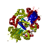
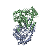
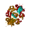
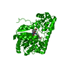
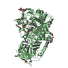
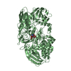
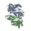
Title
Phpd with cadmium complexed with hydroethylphosphonate (HEP)
Components
PhpD
Keywords
BIOSYNTHETIC PROTEIN / Antibiotic biosynthesis / Iron dependent oxygenase
Function / homology
Function and homology information
2-hydroxyethylphosphonate dioxygenase / organic phosphonate biosynthetic process / phosphinothricin biosynthetic process / oxidoreductase activity, acting on single donors with incorporation of molecular oxygen, incorporation of two atoms of oxygen / antibiotic biosynthetic process / ferrous iron binding / protein homodimerization activity / DNA binding / identical protein binding Similarity search - Function
Resolution: 1.92→1.99 Å / Redundancy: 6.1 % / Mean I/σ(I) obs: 5.1 / Num. unique all: 2916 / Rsym value: 0.29 / % possible all: 92.5
-
Processing
Software
Name
Version
Classification
HKL-2000
datacollection
SHARP
phasing
REFMAC
5.2.0019
refinement
HKL-2000
datareduction
HKL-2000
datascaling
Refinement
Method to determine structure: SAD / Resolution: 1.92→25 Å / Cor.coef. Fo:Fc: 0.944 / Cor.coef. Fo:Fc free: 0.923 / SU B: 4.168 / SU ML: 0.123 / Cross valid method: THROUGHOUT / ESU R: 0.193 / ESU R Free: 0.172 / Stereochemistry target values: MAXIMUM LIKELIHOOD / Details: HYDROGENS HAVE BEEN ADDED IN THE RIDING POSITIONS
Rfactor
Num. reflection
% reflection
Selection details
Rfree
0.25722
1587
5.1 %
RANDOM
Rwork
0.21172
-
-
-
obs
0.21398
29741
99.16 %
-
Solvent computation
Ion probe radii: 0.8 Å / Shrinkage radii: 0.8 Å / VDW probe radii: 1.2 Å / Solvent model: BABINET MODEL WITH MASK
Displacement parameters
Biso mean: 26.621 Å2
Baniso -1
Baniso -2
Baniso -3
1-
-2.48 Å2
0 Å2
0 Å2
2-
-
3.55 Å2
0 Å2
3-
-
-
-1.07 Å2
Refinement step
Cycle: LAST / Resolution: 1.92→25 Å
Protein
Nucleic acid
Ligand
Solvent
Total
Num. atoms
3165
0
20
268
3453
Refine LS restraints
Refine-ID
Type
Dev ideal
Dev ideal target
Number
X-RAY DIFFRACTION
r_bond_refined_d
0.009
0.021
3249
X-RAY DIFFRACTION
r_angle_refined_deg
1.225
1.949
4435
X-RAY DIFFRACTION
r_dihedral_angle_1_deg
6.274
5
411
X-RAY DIFFRACTION
r_dihedral_angle_2_deg
33.648
22.759
145
X-RAY DIFFRACTION
r_dihedral_angle_3_deg
15.62
15
475
X-RAY DIFFRACTION
r_dihedral_angle_4_deg
18.165
15
29
X-RAY DIFFRACTION
r_chiral_restr
0.083
0.2
493
X-RAY DIFFRACTION
r_gen_planes_refined
0.004
0.02
2521
X-RAY DIFFRACTION
r_nbd_refined
0.209
0.2
1455
X-RAY DIFFRACTION
r_nbtor_refined
0.298
0.2
2217
X-RAY DIFFRACTION
r_xyhbond_nbd_refined
0.132
0.2
263
X-RAY DIFFRACTION
r_metal_ion_refined
0.213
0.2
5
X-RAY DIFFRACTION
r_symmetry_vdw_refined
0.236
0.2
133
X-RAY DIFFRACTION
r_symmetry_hbond_refined
0.128
0.2
37
X-RAY DIFFRACTION
r_mcbond_it
0.71
1.5
2105
X-RAY DIFFRACTION
r_mcangle_it
1.103
2
3293
X-RAY DIFFRACTION
r_scbond_it
1.844
3
1302
X-RAY DIFFRACTION
r_scangle_it
2.718
4.5
1142
LS refinement shell
Resolution: 1.92→1.97 Å / Total num. of bins used: 20
Rfactor
Num. reflection
% reflection
Rfree
0.281
106
-
Rwork
0.235
1958
-
obs
-
-
90.45 %
+
About Yorodumi
-
News
-
Feb 9, 2022. New format data for meta-information of EMDB entries
New format data for meta-information of EMDB entries
Version 3 of the EMDB header file is now the official format.
The previous official version 1.9 will be removed from the archive.
In the structure databanks used in Yorodumi, some data are registered as the other names, "COVID-19 virus" and "2019-nCoV". Here are the details of the virus and the list of structure data.
Jan 31, 2019. EMDB accession codes are about to change! (news from PDBe EMDB page)
EMDB accession codes are about to change! (news from PDBe EMDB page)
The allocation of 4 digits for EMDB accession codes will soon come to an end. Whilst these codes will remain in use, new EMDB accession codes will include an additional digit and will expand incrementally as the available range of codes is exhausted. The current 4-digit format prefixed with “EMD-” (i.e. EMD-XXXX) will advance to a 5-digit format (i.e. EMD-XXXXX), and so on. It is currently estimated that the 4-digit codes will be depleted around Spring 2019, at which point the 5-digit format will come into force.
The EM Navigator/Yorodumi systems omit the EMD- prefix.
Related info.:Q: What is EMD? / ID/Accession-code notation in Yorodumi/EM Navigator
Yorodumi is a browser for structure data from EMDB, PDB, SASBDB, etc.
This page is also the successor to EM Navigator detail page, and also detail information page/front-end page for Omokage search.
The word "yorodu" (or yorozu) is an old Japanese word meaning "ten thousand". "mi" (miru) is to see.
Related info.:EMDB / PDB / SASBDB / Comparison of 3 databanks / Yorodumi Search / Aug 31, 2016. New EM Navigator & Yorodumi / Yorodumi Papers / Jmol/JSmol / Function and homology information / Changes in new EM Navigator and Yorodumi
 Movie
Movie Controller


 Open data
 Basic information
 Components
 Keywords
 Function and homology information
 Streptomyces viridochromogenes (bacteria)
 X-RAY DIFFRACTION /
  Authors
 Citation
 Structure visualization
 Downloads & links
 Other downloads


 PDBj
PDBj


 Assembly
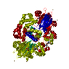
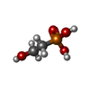
 Mass: 126.048 Da / Num. of mol.: 1 / Source method: obtained synthetically / Formula: C2H7O4P
 Mass: 112.411 Da / Num. of mol.: 13 / Source method: obtained synthetically / Formula: Cd
 Mass: 18.015 Da / Num. of mol.: 268 / Source method: isolated from a natural source / Formula: H2O
 Sample preparation
 / Beamline: 21-ID-D / Wavelength: 0.97 Å
 Processing