- PDB-3gai: Structure of a F112A variant PduO-type ATP:corrinoid adenosyltran... -
+
Open data
ID or keywords:
Loading...
-
Basic information
Entry
Database: PDB / ID: 3gai
Title
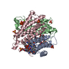
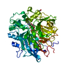
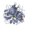
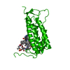
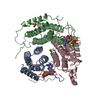
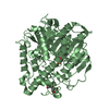
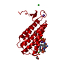
Structure of a F112A variant PduO-type ATP:corrinoid adenosyltransferase from Lactobacillus reuteri complexed with cobalamin and ATP
Components
Cobalamin adenosyltransferase PduO-like protein
Keywords
TRANSFERASE
Function / homology
Function and homology information
corrinoid adenosyltransferase / corrinoid adenosyltransferase activity / cobalamin biosynthetic process / ATP binding / metal ion binding Similarity search - Function
Journal: Biochemistry / Year: 2009 Title: Residue Phe112 of the human-type corrinoid adenosyltransferase (PduO) enzyme of Lactobacillus reuteri is critical to the formation of the four-coordinate Co(II) corrinoid substrate and to the ...Title: Residue Phe112 of the human-type corrinoid adenosyltransferase (PduO) enzyme of Lactobacillus reuteri is critical to the formation of the four-coordinate Co(II) corrinoid substrate and to the activity of the enzyme. Authors: Mera, P.E. / St Maurice, M. / Rayment, I. / Escalante-Semerena, J.C.
History
Deposition
Feb 17, 2009
Deposition site: RCSB / Processing site: RCSB
Revision 1.0
Jul 7, 2009
Provider: repository / Type: Initial release
Revision 1.1
Jul 13, 2011
Group: Non-polymer description / Version format compliance
Mass: 18.015 Da / Num. of mol.: 159 / Source method: isolated from a natural source / Formula: H2O
-
Experimental details
-
Experiment
Experiment
Method: X-RAY DIFFRACTION / Number of used crystals: 1
-
Sample preparation
Crystal
Density Matthews: 2.52 Å3/Da / Density % sol: 51.24 %
Crystal grow
Temperature: 298 K / Method: vapor diffusion, hanging drop / pH: 6 Details: anoxic, 14% PEG 8000, 0.2M potassium chloride, 0.1 M MES, 30 ug/mL E. coli FMN reductase, 50 mM NADH, 10 mM FMN, 10 mM hydoxycobalamin, 10 mM ATP, 10 mM magnesium chloride, 0.3 M sodium ...Details: anoxic, 14% PEG 8000, 0.2M potassium chloride, 0.1 M MES, 30 ug/mL E. coli FMN reductase, 50 mM NADH, 10 mM FMN, 10 mM hydoxycobalamin, 10 mM ATP, 10 mM magnesium chloride, 0.3 M sodium chloride , pH 6, VAPOR DIFFUSION, HANGING DROP, temperature 298K
-
Data collection
Diffraction
Mean temperature: 298 K
Diffraction source
Source: SYNCHROTRON / Site: APS / Beamline: 19-BM
Detector
Type: SBC-3 / Detector: CCD / Date: Aug 17, 2007
Radiation
Protocol: SINGLE WAVELENGTH / Monochromatic (M) / Laue (L): M / Scattering type: x-ray
Radiation wavelength
Relative weight: 1
Reflection
Resolution: 1.48→50 Å / Num. obs: 36190 / % possible obs: 99.7 % / Redundancy: 5.6 % / Rmerge(I) obs: 0.057 / Χ2: 2.826 / Net I/σ(I): 46.235
Reflection shell
Resolution (Å)
Redundancy (%)
Rmerge(I) obs
Num. unique all
Χ2
Diffraction-ID
% possible all
1.48-1.53
5
0.128
3618
1.121
1
98.6
1.53-1.59
5.4
0.116
3587
1.245
1
100
1.59-1.67
5.4
0.1
3644
1.509
1
100
1.67-1.75
5.5
0.089
3634
1.867
1
100
1.75-1.86
5.5
0.082
3608
2.339
1
100
1.86-2.01
5.4
0.069
3648
2.848
1
100
2.01-2.21
5.4
0.065
3631
3.906
1
100
2.21-2.53
5.4
0.059
3624
4.436
1
100
2.53-3.19
6
0.066
3573
4.945
1
98.9
3.19-50
6.5
0.049
3623
3.497
1
99.7
-
Processing
Software
Name
Version
Classification
NB
DENZO
datareduction
SCALEPACK
datascaling
REFMAC
5.2.0005
refinement
PDB_EXTRACT
3.006
dataextraction
Refinement
Resolution: 1.48→30 Å / Cor.coef. Fo:Fc: 0.973 / Cor.coef. Fo:Fc free: 0.969 / Occupancy max: 1 / Occupancy min: 0 / SU B: 0.867 / SU ML: 0.035 / Cross valid method: THROUGHOUT / σ(F): 0 / ESU R: 0.061 / ESU R Free: 0.061 / Stereochemistry target values: MAXIMUM LIKELIHOOD / Details: HYDROGENS HAVE BEEN ADDED IN THE RIDING POSITIONS
Rfactor
Num. reflection
% reflection
Selection details
Rfree
0.174
1804
5 %
RANDOM
Rwork
0.156
-
-
-
obs
0.157
36185
99.69 %
-
Solvent computation
Ion probe radii: 0.8 Å / Shrinkage radii: 0.8 Å / VDW probe radii: 1.2 Å / Solvent model: MASK
In the structure databanks used in Yorodumi, some data are registered as the other names, "COVID-19 virus" and "2019-nCoV". Here are the details of the virus and the list of structure data.
Jan 31, 2019. EMDB accession codes are about to change! (news from PDBe EMDB page)
EMDB accession codes are about to change! (news from PDBe EMDB page)
The allocation of 4 digits for EMDB accession codes will soon come to an end. Whilst these codes will remain in use, new EMDB accession codes will include an additional digit and will expand incrementally as the available range of codes is exhausted. The current 4-digit format prefixed with “EMD-” (i.e. EMD-XXXX) will advance to a 5-digit format (i.e. EMD-XXXXX), and so on. It is currently estimated that the 4-digit codes will be depleted around Spring 2019, at which point the 5-digit format will come into force.
The EM Navigator/Yorodumi systems omit the EMD- prefix.
Related info.:Q: What is EMD? / ID/Accession-code notation in Yorodumi/EM Navigator
Yorodumi is a browser for structure data from EMDB, PDB, SASBDB, etc.
This page is also the successor to EM Navigator detail page, and also detail information page/front-end page for Omokage search.
The word "yorodu" (or yorozu) is an old Japanese word meaning "ten thousand". "mi" (miru) is to see.
Related info.:EMDB / PDB / SASBDB / Comparison of 3 databanks / Yorodumi Search / Aug 31, 2016. New EM Navigator & Yorodumi / Yorodumi Papers / Jmol/JSmol / Function and homology information / Changes in new EM Navigator and Yorodumi
 Movie
Movie Controller

 Yorodumi
 Open data
 Basic information
 Components
 Keywords
 Function and homology information
 Lactobacillus reuteri (bacteria)
 X-RAY DIFFRACTION /
  Authors
 Citation
 Structure visualization
 Downloads & links
 Other downloads
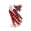


 PDBj
PDBj Assembly
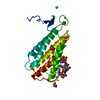


 Mass: 507.181 Da / Num. of mol.: 1 / Source method: obtained synthetically / Formula: C10H16N5O13P3 / Comment: ATP, energy-carrying molecule*YM
 Mass: 1330.356 Da / Num. of mol.: 1 / Source method: obtained synthetically / Formula: C62H89CoN13O14P
 Mass: 24.305 Da / Num. of mol.: 1 / Source method: obtained synthetically / Formula: Mg
 Mass: 39.098 Da / Num. of mol.: 2 / Source method: obtained synthetically / Formula: K
 Mass: 35.453 Da / Num. of mol.: 2 / Source method: obtained synthetically / Formula: Cl
 Mass: 92.094 Da / Num. of mol.: 3 / Source method: obtained synthetically / Formula: C3H8O3
 Sample preparation
 / Beamline: 19-BM
 Processing