 Movie
Movie Controller

[English] 日本語
 Yorodumi
- PDB-3gaa: The crystal structure of the protein with unknown function from T... -
+ Open data

- Basic information
| Entry | Database: PDB / ID: 3gaa | ||||||
|---|---|---|---|---|---|---|---|
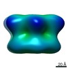
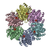
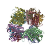
| Title | The crystal structure of the protein with unknown function from Thermoplasma acidophilum | ||||||
 Components Components | uncharacterized protein Ta1441 | ||||||
 Keywords Keywords | structural genomics / unknown function / The protein with unknown function from Thermoplasma acidophilum / PSI / MCSG / Protein Structure Initiative / Midwest Center for Structural Genomics | ||||||
| Function / homology | PAC-like subunit / Proteasome assembly chaperone 2 / Proteasome assembly chaperone 2 superfamily / PAC2 family / Rossmann fold / 3-Layer(aba) Sandwich / Alpha Beta / Proteasome assembly chaperone family protein Function and homology information Function and homology information | ||||||
| Biological species |   Thermoplasma acidophilum (acidophilic) Thermoplasma acidophilum (acidophilic) | ||||||
| Method |  X-RAY DIFFRACTION / SYNCHROTRON / SAD / Resolution: 2.7 Å X-RAY DIFFRACTION / SYNCHROTRON / SAD / Resolution: 2.7 Å | ||||||
 Authors Authors | Zhang, R. / Borovilos, M. / Clancy, S. / Joachimiak, A. / Midwest Center for Structural Genomics (MCSG) | ||||||
 Citation Citation | Journal: To be Published Title: The crystal structure of the protein with unknown function from Thermoplasma acidophilum Authors: Zhang, R. / Borovilos, M. / Clancy, S. / Joachimiak, A. | ||||||
| History |
|
- Structure visualization
| Structure viewer | Molecule: MolmilJmol/JSmol |
|---|
- Downloads & links
-Download
| PDBx/mmCIF format | 3gaa.cif.gz | 231.8 KB | Display | PDBx/mmCIF format |
|---|---|---|---|---|
| PDB format | pdb3gaa.ent.gz | 190 KB | Display | PDB format |
| PDBx/mmJSON format | 3gaa.json.gz | Tree view | PDBx/mmJSON format | |
| Others |  Other downloads Other downloads |
-Validation report
| Arichive directory | https://data.pdbj.org/pub/pdb/validation_reports/ga/3gaaftp://data.pdbj.org/pub/pdb/validation_reports/ga/3gaa | HTTPS FTP |
|---|
-Related structure data
| Similar structure data | |
|---|---|
| Other databases |


-Links
 PDBj
PDBj- Assembly
| Deposited unit | 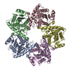
| ||||||||
|---|---|---|---|---|---|---|---|---|---|
| 1 | 
| ||||||||
| Unit cell |
| ||||||||
| Details | This protein existed as decamer. The second part of the decamer is generated by the axis: y,x,-z+1 |
-Components
| #1: Protein | Mass: 27588.037 Da / Num. of mol.: 5 Source method: isolated from a genetically manipulated source Source: (gene. exp.) Thermoplasma acidophilum (acidophilic) / Strain: DSM1728 / Gene: GI:10640783, Ta1441 / Plasmid: pMCSG7 / Production host:  #2: Water | ChemComp-HOH / |  Mass: 18.015 Da / Num. of mol.: 35 / Source method: isolated from a natural source / Formula: H2O Mass: 18.015 Da / Num. of mol.: 35 / Source method: isolated from a natural source / Formula: H2O |
|---|
-Experimental details
-Experiment
| Experiment | Method: X-RAY DIFFRACTION / Number of used crystals: 1 |
|---|
- Sample preparation
| Crystal | Density Matthews: 3.09 Å3/Da / Density % sol: 60.15 % |
|---|---|
| Crystal grow | Temperature: 289 K / Method: vapor diffusion, sitting drop / pH: 6.5 Details: 25% PEG4000,0.1M Mes, pH 6.5, VAPOR DIFFUSION, SITTING DROP, temperature 289K |
-Data collection
| Diffraction | Mean temperature: 100 K |
|---|---|
| Diffraction source | Source: SYNCHROTRON / Site: APS  / Beamline: 19-ID / Wavelength: 0.9794 Å / Beamline: 19-ID / Wavelength: 0.9794 Å |
| Detector | Type: ADSC QUANTUM 315 / Detector: CCD / Date: Sep 19, 2006 / Details: mirrors |
| Radiation | Monochromator: Si 111 channel / Protocol: SINGLE WAVELENGTH / Monochromatic (M) / Laue (L): M / Scattering type: x-ray |
| Radiation wavelength | Wavelength: 0.9794 Å / Relative weight: 1 |
| Reflection | Resolution: 2.7→101.02 Å / Num. all: 44574 / Num. obs: 44396 / % possible obs: 99.6 % / Observed criterion σ(F): 1 / Observed criterion σ(I): 1 / Redundancy: 9.7 % / Rmerge(I) obs: 0.14 / Net I/σ(I): 17.5 |
| Reflection shell | Resolution: 2.7→2.768 Å / Redundancy: 6.3 % / Rmerge(I) obs: 0.67 / Mean I/σ(I) obs: 1.17 / Num. unique all: 3446 / % possible all: 97.53 |
- Processing
| Software |
| |||||||||||||||||||||||||||||||||||||||||||||||||||||||||||||||||||||||||||||||||||||
|---|---|---|---|---|---|---|---|---|---|---|---|---|---|---|---|---|---|---|---|---|---|---|---|---|---|---|---|---|---|---|---|---|---|---|---|---|---|---|---|---|---|---|---|---|---|---|---|---|---|---|---|---|---|---|---|---|---|---|---|---|---|---|---|---|---|---|---|---|---|---|---|---|---|---|---|---|---|---|---|---|---|---|---|---|---|---|
| Refinement | Method to determine structure: SAD / Resolution: 2.7→101.02 Å / Cor.coef. Fo:Fc: 0.954 / Cor.coef. Fo:Fc free: 0.924 / SU B: 28.768 / SU ML: 0.255 / TLS residual ADP flag: LIKELY RESIDUAL / Cross valid method: THROUGHOUT / σ(F): 1 / σ(I): 0 / ESU R: 0.618 / ESU R Free: 0.322 Stereochemistry target values: MAXIMUM LIKELIHOOD WITH PHASES Details: HYDROGENS HAVE BEEN ADDED IN THE RIDING POSITIONS
| |||||||||||||||||||||||||||||||||||||||||||||||||||||||||||||||||||||||||||||||||||||
| Solvent computation | Ion probe radii: 0.8 Å / Shrinkage radii: 0.8 Å / VDW probe radii: 1.2 Å / Solvent model: MASK | |||||||||||||||||||||||||||||||||||||||||||||||||||||||||||||||||||||||||||||||||||||
| Displacement parameters | Biso mean: 40.639 Å2
| |||||||||||||||||||||||||||||||||||||||||||||||||||||||||||||||||||||||||||||||||||||
| Refinement step | Cycle: LAST / Resolution: 2.7→101.02 Å
| |||||||||||||||||||||||||||||||||||||||||||||||||||||||||||||||||||||||||||||||||||||
| Refine LS restraints |
| |||||||||||||||||||||||||||||||||||||||||||||||||||||||||||||||||||||||||||||||||||||
| LS refinement shell | Resolution: 2.698→2.768 Å / Total num. of bins used: 20
| |||||||||||||||||||||||||||||||||||||||||||||||||||||||||||||||||||||||||||||||||||||
| Refinement TLS params. | Method: refined / Origin x: 48.853 Å / Origin y: 87.215 Å / Origin z: 109.259 Å
| |||||||||||||||||||||||||||||||||||||||||||||||||||||||||||||||||||||||||||||||||||||
| Refinement TLS group |
|
