Mass: 18.015 Da / Num. of mol.: 532 / Source method: isolated from a natural source / Formula: H2O
Has protein modification
Y
Sequence details
THE CONSTRUCT WAS EXPRESSED WITH A PURIFICATION TAG MGSDKIHHHHHHENLYFQG. THE TAG WAS REMOVED WITH ...THE CONSTRUCT WAS EXPRESSED WITH A PURIFICATION TAG MGSDKIHHHHHHENLYFQG. THE TAG WAS REMOVED WITH TEV PROTEASE LEAVING ONLY A GLYCINE (0) FOLLOWED BY THE TARGET SEQUENCE.
-
Experimental details
-
Experiment
Experiment
Method: X-RAY DIFFRACTION / Number of used crystals: 1
-
Sample preparation
Crystal
Density Matthews: 2.47 Å3/Da / Density % sol: 50.22 %
Crystal grow
Temperature: 277 K / Method: vapor diffusion, sitting drop / pH: 6.5 Details: NANODROP, 0.20M MgCl2, 10.0% PEG 3000, 0.1M Cacodylate pH 6.5, VAPOR DIFFUSION, SITTING DROP, temperature 277K
Type: MARMOSAIC 325 mm CCD / Detector: CCD / Date: Nov 15, 2008 / Details: Flat mirror (vertical focusing)
Radiation
Monochromator: Single crystal Si(111) bent (horizontal focusing) Protocol: MAD / Monochromatic (M) / Laue (L): M / Scattering type: x-ray
Radiation wavelength
ID
Wavelength (Å)
Relative weight
1
0.91162
1
2
0.97821
1
3
0.97879
1
Reflection
Resolution: 1.8→29.285 Å / Num. obs: 52108 / % possible obs: 98.8 % / Redundancy: 3.7 % / Biso Wilson estimate: 19.743 Å2 / Rmerge(I) obs: 0.108 / Rsym value: 0.108 / Net I/σ(I): 4.81
Reflection shell
Diffraction-ID: 1
Resolution (Å)
Redundancy (%)
Rmerge(I) obs
Mean I/σ(I) obs
Num. measured all
Num. unique all
Rsym value
% possible all
1.8-1.85
3.7
0.753
1
13918
3761
0.753
98
1.85-1.9
3.7
0.646
1.2
13717
3696
0.646
98.1
1.9-1.95
3.7
0.52
1.5
13253
3573
0.52
98.1
1.95-2.01
3.7
0.429
1.8
12938
3490
0.429
98.6
2.01-2.08
3.7
0.332
2.3
12569
3394
0.332
98.5
2.08-2.15
3.7
0.279
2.7
12176
3274
0.279
98.3
2.15-2.23
3.7
0.239
3.1
11925
3202
0.239
98.9
2.23-2.32
3.7
0.196
3.8
11427
3061
0.196
98.7
2.32-2.43
3.7
0.168
4.3
10946
2935
0.168
99
2.43-2.55
3.7
0.154
4.7
10625
2848
0.154
98.9
2.55-2.68
3.7
0.129
5.5
10075
2712
0.129
99.3
2.68-2.85
3.7
0.107
6.3
9496
2559
0.107
99
2.85-3.04
3.7
0.086
6.8
8808
2389
0.086
99.3
3.04-3.29
3.7
0.072
7.8
8339
2278
0.072
99.3
3.29-3.6
3.6
0.062
8.1
7605
2093
0.062
99.4
3.6-4.02
3.6
0.056
7.6
6884
1900
0.056
99.3
4.02-4.65
3.6
0.049
9.4
6108
1695
0.049
99.5
4.65-5.69
3.5
0.049
9.7
5127
1447
0.049
99.6
5.69-8.05
3.5
0.048
12.8
3962
1147
0.048
99.7
8.05-29.285
3.2
0.045
11.6
2125
654
0.045
96.9
-
Phasing
Phasing
Method: MAD
-
Processing
Software
Name
Version
Classification
NB
REFMAC
5.2.0019
refinement
PHENIX
refinement
SHELX
phasing
MolProbity
3beta29
modelbuilding
SCALA
3.2.5
datascaling
PDB_EXTRACT
3.006
dataextraction
MAR345
CCD
datacollection
MOSFLM
datareduction
SHELXD
phasing
autoSHARP
phasing
Refinement
Method to determine structure: MAD / Resolution: 1.8→29.285 Å / Cor.coef. Fo:Fc: 0.953 / Cor.coef. Fo:Fc free: 0.929 / Occupancy max: 1 / Occupancy min: 0.2 / SU B: 4.731 / SU ML: 0.135 / Cross valid method: THROUGHOUT / σ(F): 0 / ESU R: 0.143 / ESU R Free: 0.134 Stereochemistry target values: MAXIMUM LIKELIHOOD WITH PHASES Details: 1. HYDROGENS HAVE BEEN ADDED IN THE RIDING POSITIONS. 2. A MET-INHIBITION PROTOCOL WAS USED FOR SELENOMETHIONINE INCORPORATION DURING PROTEIN EXPRESSION. THE OCCUPANCY OF THE SE ATOMS IN THE ...Details: 1. HYDROGENS HAVE BEEN ADDED IN THE RIDING POSITIONS. 2. A MET-INHIBITION PROTOCOL WAS USED FOR SELENOMETHIONINE INCORPORATION DURING PROTEIN EXPRESSION. THE OCCUPANCY OF THE SE ATOMS IN THE MSE RESIDUES WAS REDUCED TO 0.75 TO ACCOUNT FOR THE REDUCED SCATTERING POWER DUE TO PARTIAL S-MET INCORPORATION. 3. CACODYLATE ANION, NA ION AND ETHYLENE GLYCOL FROM CRYSTALLIZATION AND CRYOPROTECTANT ARE MODELED INTO THIS STRUCTURE, RESPECTIVELY.
Rfactor
Num. reflection
% reflection
Selection details
Rfree
0.239
2652
5.1 %
RANDOM
Rwork
0.203
-
-
-
obs
0.205
52068
98.49 %
-
Solvent computation
Ion probe radii: 0.8 Å / Shrinkage radii: 0.8 Å / VDW probe radii: 1.2 Å / Solvent model: BABINET MODEL WITH MASK
In the structure databanks used in Yorodumi, some data are registered as the other names, "COVID-19 virus" and "2019-nCoV". Here are the details of the virus and the list of structure data.
Jan 31, 2019. EMDB accession codes are about to change! (news from PDBe EMDB page)
EMDB accession codes are about to change! (news from PDBe EMDB page)
The allocation of 4 digits for EMDB accession codes will soon come to an end. Whilst these codes will remain in use, new EMDB accession codes will include an additional digit and will expand incrementally as the available range of codes is exhausted. The current 4-digit format prefixed with “EMD-” (i.e. EMD-XXXX) will advance to a 5-digit format (i.e. EMD-XXXXX), and so on. It is currently estimated that the 4-digit codes will be depleted around Spring 2019, at which point the 5-digit format will come into force.
The EM Navigator/Yorodumi systems omit the EMD- prefix.
Related info.:Q: What is EMD? / ID/Accession-code notation in Yorodumi/EM Navigator
Yorodumi is a browser for structure data from EMDB, PDB, SASBDB, etc.
This page is also the successor to EM Navigator detail page, and also detail information page/front-end page for Omokage search.
The word "yorodu" (or yorozu) is an old Japanese word meaning "ten thousand". "mi" (miru) is to see.
Related info.:EMDB / PDB / SASBDB / Comparison of 3 databanks / Yorodumi Search / Aug 31, 2016. New EM Navigator & Yorodumi / Yorodumi Papers / Jmol/JSmol / Function and homology information / Changes in new EM Navigator and Yorodumi
 Movie
Movie Controller

 Yorodumi
 Open data
 Basic information
 Components
 Keywords
 Function and homology information
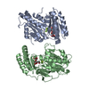
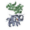
 Streptococcus suis (bacteria)
 X-RAY DIFFRACTION /
  Authors
 Citation
 Structure visualization
 Downloads & links
 Other downloads
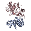


 PDBj
PDBj


 Assembly

 Mass: 22.990 Da / Num. of mol.: 1 / Source method: obtained synthetically / Formula: Na
 Mass: 136.989 Da / Num. of mol.: 1 / Source method: obtained synthetically / Formula: C2H6AsO2
 Mass: 62.068 Da / Num. of mol.: 6 / Source method: obtained synthetically / Formula: C2H6O2
 Mass: 18.015 Da / Num. of mol.: 532 / Source method: isolated from a natural source / Formula: H2O
 Sample preparation
 / Beamline: BL11-1 / Wavelength: 0.91162, 0.97821, 0.97879
 Processing