 Movie
Movie Controller

[English] 日本語
 Yorodumi
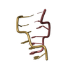
- PDB-3g2a: Crystal structure of d(CACGCG).d(CGCGTG) grown in presence of 1mM... -
+ Open data

- Basic information
| Entry | Database: PDB / ID: 3g2a | ||||||
|---|---|---|---|---|---|---|---|
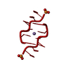
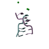
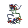
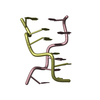
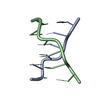
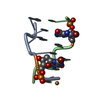
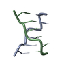
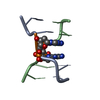
| Title | Crystal structure of d(CACGCG).d(CGCGTG) grown in presence of 1mM MnCl2 | ||||||
 Components Components |
| ||||||
 Keywords Keywords | DNA / double helical Z form DNA | ||||||
| Function / homology | DNA Function and homology information Function and homology information | ||||||
| Method |  X-RAY DIFFRACTION / MOLECULAR REPLACEMENT / Resolution: 2 Å X-RAY DIFFRACTION / MOLECULAR REPLACEMENT / Resolution: 2 Å | ||||||
 Authors Authors | Venkadesh, S. / Mandal, P.K. / Kannan, R. / Gautham, N. | ||||||
 Citation Citation | Journal: To be Published Title: Crystal Structures of d(CACGCG).d(CGCGTG) cocrystallized with MnCl2 Authors: Venkadesh, S. / Mandal, P.K. / Gautham, N. | ||||||
| History |
|
- Structure visualization
| Structure viewer | Molecule: MolmilJmol/JSmol |
|---|
- Downloads & links
-Download
| PDBx/mmCIF format | 3g2a.cif.gz | 16.8 KB | Display | PDBx/mmCIF format |
|---|---|---|---|---|
| PDB format | pdb3g2a.ent.gz | 10.3 KB | Display | PDB format |
| PDBx/mmJSON format | 3g2a.json.gz | Tree view | PDBx/mmJSON format | |
| Others |  Other downloads Other downloads |
-Validation report
| Arichive directory | https://data.pdbj.org/pub/pdb/validation_reports/g2/3g2aftp://data.pdbj.org/pub/pdb/validation_reports/g2/3g2a | HTTPS FTP |
|---|
-Related structure data
| Related structure data | |
|---|---|
| Similar structure data |


-Links
 PDBj
PDBj

- Assembly
| Deposited unit | 
| ||||||||
|---|---|---|---|---|---|---|---|---|---|
| 1 |
| ||||||||
| Unit cell |
|
-Components
| #1: DNA chain | Mass: 1794.206 Da / Num. of mol.: 1 / Source method: obtained synthetically |
|---|---|
| #2: DNA chain | Mass: 1825.216 Da / Num. of mol.: 1 / Source method: obtained synthetically |
| #3: Water | ChemComp-HOH /  Mass: 18.015 Da / Num. of mol.: 24 / Source method: isolated from a natural source / Formula: H2O Mass: 18.015 Da / Num. of mol.: 24 / Source method: isolated from a natural source / Formula: H2O |
-Experimental details
-Experiment
| Experiment | Method: X-RAY DIFFRACTION / Number of used crystals: 1 |
|---|
- Sample preparation
| Crystal | Density Matthews: 1.7 Å3/Da / Density % sol: 27.78 % | ||||||||||||||||||||||||||||||||
|---|---|---|---|---|---|---|---|---|---|---|---|---|---|---|---|---|---|---|---|---|---|---|---|---|---|---|---|---|---|---|---|---|---|
| Crystal grow | Temperature: 293 K / Method: vapor diffusion, hanging drop / pH: 6.99 Details: 50mM Sodium Cacodylate, 1mM MnCl2, 1mM Spermine, 50% Methyl pentane diol, pH 6.99, VAPOR DIFFUSION, HANGING DROP, temperature 293K | ||||||||||||||||||||||||||||||||
| Components of the solutions |
|
-Data collection
| Diffraction | Mean temperature: 293 K |
|---|---|
| Diffraction source | Source: ROTATING ANODE / Type: RIGAKU / Wavelength: 1.54 Å |
| Detector | Type: MARRESEARCH / Detector: IMAGE PLATE / Date: Sep 9, 2008 |
| Radiation | Monochromator: mirror / Protocol: SINGLE WAVELENGTH / Monochromatic (M) / Laue (L): M / Scattering type: x-ray |
| Radiation wavelength | Wavelength: 1.54 Å / Relative weight: 1 |
| Reflection | Resolution: 2→15 Å / Num. all: 1914 / Num. obs: 1819 / % possible obs: 95.2 % / Redundancy: 4.56 % / Biso Wilson estimate: 27.1 Å2 / Rmerge(I) obs: 0.105 / Rsym value: 0.0896 / Net I/σ(I): 4.1 |
| Reflection shell | Resolution: 2→2.07 Å / Redundancy: 4.69 % / Rmerge(I) obs: 0.2886 / Mean I/σ(I) obs: 1.2 / Num. unique all: 172 / Rsym value: 0.258 / % possible all: 99.4 |
- Processing
| Software |
| ||||||||||||||||||||||||||||||||||||||||||||||||||
|---|---|---|---|---|---|---|---|---|---|---|---|---|---|---|---|---|---|---|---|---|---|---|---|---|---|---|---|---|---|---|---|---|---|---|---|---|---|---|---|---|---|---|---|---|---|---|---|---|---|---|---|
| Refinement | Method to determine structure: MOLECULAR REPLACEMENT / Resolution: 2→15 Å / Cor.coef. Fo:Fc: 0.905 / Cor.coef. Fo:Fc free: 0.965 / SU B: 5.557 / SU ML: 0.146 / Isotropic thermal model: Isotropic / Cross valid method: THROUGHOUT / ESU R: 0.263 / ESU R Free: 0.195 / Stereochemistry target values: MAXIMUM LIKELIHOOD
| ||||||||||||||||||||||||||||||||||||||||||||||||||
| Solvent computation | Ion probe radii: 0.8 Å / Shrinkage radii: 0.8 Å / VDW probe radii: 1.2 Å / Solvent model: MASK | ||||||||||||||||||||||||||||||||||||||||||||||||||
| Displacement parameters | Biso mean: 12.664 Å2
| ||||||||||||||||||||||||||||||||||||||||||||||||||
| Refinement step | Cycle: LAST / Resolution: 2→15 Å
| ||||||||||||||||||||||||||||||||||||||||||||||||||
| Refine LS restraints |
| ||||||||||||||||||||||||||||||||||||||||||||||||||
| LS refinement shell | Resolution: 2→2.051 Å / Total num. of bins used: 20
|
