 Movie
Movie Controller


+ Open data

- Basic information
| Entry | Database: PDB / ID: 3fva | ||||||
|---|---|---|---|---|---|---|---|
| Title | NNQNTF segment from elk prion | ||||||
 Components Components | Major prion protein | ||||||
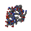
 Keywords Keywords | PROTEIN FIBRIL / amyloid-like protofibril / Cell membrane / Glycoprotein / Golgi apparatus / GPI-anchor / Lipoprotein / Membrane / Prion | ||||||
| Function / homology |  Function and homology information Function and homology informationside of membrane / protein homooligomerization / copper ion binding / Golgi apparatus / identical protein binding / plasma membrane Similarity search - Function | ||||||
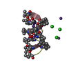
| Method |  X-RAY DIFFRACTION / SYNCHROTRON / MOLECULAR REPLACEMENT / molecular replacement / Resolution: 1.458 Å X-RAY DIFFRACTION / SYNCHROTRON / MOLECULAR REPLACEMENT / molecular replacement / Resolution: 1.458 Å | ||||||
 Authors Authors | Apostol, M.I. / Eisenberg, D. | ||||||
 Citation Citation | Journal: Nat.Struct.Mol.Biol. / Year: 2009 Title: Molecular mechanisms for protein-encoded inheritance. Authors: Wiltzius, J.J. / Landau, M. / Nelson, R. / Sawaya, M.R. / Apostol, M.I. / Goldschmidt, L. / Soriaga, A.B. / Cascio, D. / Rajashankar, K. / Eisenberg, D. | ||||||
| History |
|
- Structure visualization
| Structure viewer | Molecule: MolmilJmol/JSmol |
|---|
- Downloads & links
-Download
| PDBx/mmCIF format | 3fva.cif.gz | 8.5 KB | Display | PDBx/mmCIF format |
|---|---|---|---|---|
| PDB format | pdb3fva.ent.gz | 5.1 KB | Display | PDB format |
| PDBx/mmJSON format | 3fva.json.gz | Tree view | PDBx/mmJSON format | |
| Others |  Other downloads Other downloads |
-Validation report
| Arichive directory | https://data.pdbj.org/pub/pdb/validation_reports/fv/3fvaftp://data.pdbj.org/pub/pdb/validation_reports/fv/3fva | HTTPS FTP |
|---|
-Related structure data
| Related structure data |  3fodC  3fpoC  3fr1C  3fthC  3ftkC  3ftlC  3ftrC 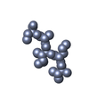 4np8C C: citing same article ( |
|---|---|
| Similar structure data |


-Links
 PDBj
PDBj

- Assembly
| Deposited unit | 
| ||||||||
|---|---|---|---|---|---|---|---|---|---|
| 1 | x 6
| ||||||||
| Unit cell |
| ||||||||
| Details | Authors state that the biological unit is an indefinitely long pair of sheets (a protofibril). One sheet is constructed from chain A (X,Y,Z) and unit cell translations along b cell dimension (e.g. X,Y+1,Z; X,Y-1,Z). The second sheet is constructed from crystallographic symmetry operator -x-1,y+1/2,-z-1 and unit cell translations along the b dimension (e.g. -x-1,y+3/2,-z-1). There is an additional polymorph of the biological unit (also a pair of beta sheets). One sheet is constructed from chain A (X,Y,Z) and unit cell translations along b cell dimension (e.g. X,Y+1,Z; X,Y-1,Z). The second sheet is constructed from crystallographic symmetry operator -x,y+1/2,-z-1 and unit cell translations along the b dimension (e.g. -x,y+3/2,-z-1). |
-Components
| #1: Protein/peptide | Mass: 736.730 Da / Num. of mol.: 1 / Fragment: NNQNTF (residues 173-178) / Source method: obtained synthetically / Details: NNQNTF (residues 173-178) from elk prion protein / References: UniProt: P67986 |
|---|---|
| #2: Water | ChemComp-HOH /  Mass: 18.015 Da / Num. of mol.: 2 / Source method: isolated from a natural source / Formula: H2O Mass: 18.015 Da / Num. of mol.: 2 / Source method: isolated from a natural source / Formula: H2O |
-Experimental details
-Experiment
| Experiment | Method: X-RAY DIFFRACTION / Number of used crystals: 1 |
|---|
- Sample preparation
| Crystal | Density Matthews: 1.248 Å3/Da / Density % sol: 1.417 % |
|---|---|
| Crystal grow | Temperature: 298 K / Method: vapor diffusion, hanging drop / pH: 8.5 Details: 0.2M Ammonium Sulfate, 0.1M Tris pH 8.5, 25% PEG 3350, vapor diffusion, hanging drop, temperature 298K |
-Data collection
| Diffraction | Mean temperature: 100 K | ||||||||||||||||||||||||||||||||||||||||||||||||||||||||||||||||||
|---|---|---|---|---|---|---|---|---|---|---|---|---|---|---|---|---|---|---|---|---|---|---|---|---|---|---|---|---|---|---|---|---|---|---|---|---|---|---|---|---|---|---|---|---|---|---|---|---|---|---|---|---|---|---|---|---|---|---|---|---|---|---|---|---|---|---|---|
| Diffraction source | Source: SYNCHROTRON / Site: ESRF  / Beamline: ID13 / Wavelength: 0.946496 Å / Beamline: ID13 / Wavelength: 0.946496 Å | ||||||||||||||||||||||||||||||||||||||||||||||||||||||||||||||||||
| Detector | Type: MARRESEARCH / Detector: CCD / Date: May 13, 2006 | ||||||||||||||||||||||||||||||||||||||||||||||||||||||||||||||||||
| Radiation | Protocol: SINGLE WAVELENGTH / Monochromatic (M) / Laue (L): M / Scattering type: x-ray | ||||||||||||||||||||||||||||||||||||||||||||||||||||||||||||||||||
| Radiation wavelength | Wavelength: 0.946496 Å / Relative weight: 1 | ||||||||||||||||||||||||||||||||||||||||||||||||||||||||||||||||||
| Reflection | Resolution: 1.45→30 Å / Num. obs: 750 / % possible obs: 98.7 % / Observed criterion σ(I): -3 / Redundancy: 5.4 % / Biso Wilson estimate: 10.5 Å2 / Rmerge(I) obs: 0.137 / Χ2: 1.031 / Net I/σ(I): 7.197 | ||||||||||||||||||||||||||||||||||||||||||||||||||||||||||||||||||
| Reflection shell |
|
-Phasing
| Phasing | Method: molecular replacement |
|---|
- Processing
| Software |
| |||||||||||||||||||||||||||||||||||||||||||||||||||||||||||||||||||||||||||
|---|---|---|---|---|---|---|---|---|---|---|---|---|---|---|---|---|---|---|---|---|---|---|---|---|---|---|---|---|---|---|---|---|---|---|---|---|---|---|---|---|---|---|---|---|---|---|---|---|---|---|---|---|---|---|---|---|---|---|---|---|---|---|---|---|---|---|---|---|---|---|---|---|---|---|---|---|
| Refinement | Method to determine structure: MOLECULAR REPLACEMENT / Resolution: 1.458→21.03 Å / Cor.coef. Fo:Fc: 0.96 / Cor.coef. Fo:Fc free: 0.974 / WRfactor Rfree: 0.187 / WRfactor Rwork: 0.193 / Occupancy max: 1 / Occupancy min: 1 / FOM work R set: 0.914 / SU B: 0.981 / SU ML: 0.038 / SU R Cruickshank DPI: 0.08 / SU Rfree: 0.072 / Cross valid method: THROUGHOUT / σ(F): 0 / ESU R: 0.08 / ESU R Free: 0.072 / Stereochemistry target values: MAXIMUM LIKELIHOOD / Details: HYDROGENS HAVE BEEN ADDED IN THE RIDING POSITIONS
| |||||||||||||||||||||||||||||||||||||||||||||||||||||||||||||||||||||||||||
| Solvent computation | Ion probe radii: 0.8 Å / Shrinkage radii: 0.8 Å / VDW probe radii: 1.4 Å / Solvent model: MASK | |||||||||||||||||||||||||||||||||||||||||||||||||||||||||||||||||||||||||||
| Displacement parameters | Biso max: 11.11 Å2 / Biso mean: 0.702 Å2 / Biso min: 0 Å2
| |||||||||||||||||||||||||||||||||||||||||||||||||||||||||||||||||||||||||||
| Refinement step | Cycle: LAST / Resolution: 1.458→21.03 Å /
| |||||||||||||||||||||||||||||||||||||||||||||||||||||||||||||||||||||||||||
| Refine LS restraints |
| |||||||||||||||||||||||||||||||||||||||||||||||||||||||||||||||||||||||||||
| LS refinement shell | Resolution: 1.458→1.496 Å / Total num. of bins used: 20
|
