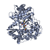
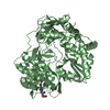
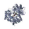
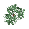
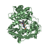
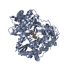
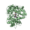
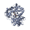
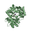
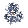
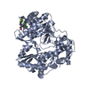
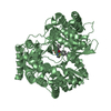
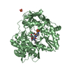
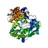
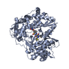
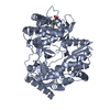
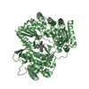
[English] 日本語
Yorodumi
- PDB-3fql: Hepatitis C virus polymerase NS5B (CON1 1-570) with HCV-796 inhibitor -

+
Open data


ID or keywords:

Loading...

-
Basic information

Entry
Database: PDB / ID: 3fql
TitleHepatitis C virus polymerase NS5B (CON1 1-570) with HCV-796 inhibitor
ComponentsRNA-directed RNA polymeraseRNA-dependent RNA polymerase
KeywordsTRANSFERASE / HCV / HEPATITIS / NS5B / RNA-DEPENDENT RNA POLYMERASE / HCV-796
Function / homology
Function and homology information


positive stranded viral RNA replication / hepacivirin / host cell mitochondrial membrane / host cell lipid droplet / symbiont-mediated suppression of host TRAF-mediated signal transduction / transformation of host cell by virus / symbiont-mediated perturbation of host cell cycle G1/S transition checkpoint / symbiont-mediated suppression of host JAK-STAT cascade via inhibition of STAT1 activity / symbiont-mediated suppression of host cytoplasmic pattern recognition receptor signaling pathway via inhibition of MAVS activity / viral genome replication ...positive stranded viral RNA replication / hepacivirin / host cell mitochondrial membrane / host cell lipid droplet / symbiont-mediated suppression of host TRAF-mediated signal transduction / transformation of host cell by virus / symbiont-mediated perturbation of host cell cycle G1/S transition checkpoint / symbiont-mediated suppression of host JAK-STAT cascade via inhibition of STAT1 activity / symbiont-mediated suppression of host cytoplasmic pattern recognition receptor signaling pathway via inhibition of MAVS activity / viral genome replication / SH3 domain binding / : / nucleoside-triphosphate phosphatase / protein complex oligomerization / monoatomic ion channel activity / clathrin-dependent endocytosis of virus by host cell / viral nucleocapsid / Hydrolases; Acting on peptide bonds (peptidases); Cysteine endopeptidases / RNA helicase activity / host cell endoplasmic reticulum membrane / host cell perinuclear region of cytoplasm / RNA helicase / induction by virus of host autophagy / ribonucleoprotein complex / RNA-directed RNA polymerase / viral RNA genome replication / cysteine-type endopeptidase activity / RNA-dependent RNA polymerase activity / serine-type endopeptidase activity / fusion of virus membrane with host endosome membrane / viral envelope / symbiont-mediated suppression of host type I interferon-mediated signaling pathway / host cell nucleus / virion attachment to host cell / host cell plasma membrane / virion membrane / structural molecule activity / ATP hydrolysis activity / protein-containing complex / proteolysis / RNA binding / zinc ion binding / ATP binding / membrane / identical protein binding
Similarity search - Function
Hepatitis C virus core protein, chain A superfamily / Hepatitus C virus, Non-structural 5a protein, C-terminal / Hepatitis C virus NS5A, 1B domain superfamily / Hepatitis C virus non-structural protein NS2, C-terminal domain / Hepatitis C virus non-structural protein NS2, N-terminal domain / Hepatitis C virus non-structural protein NS2 / HCV NS5a protein C-terminal region / Hepatitis C virus, Non-structural protein NS4b / Hepatitis C virus, Core protein, N-terminal / Hepatitis C virus non-structural protein NS4b ...Hepatitis C virus core protein, chain A superfamily / Hepatitus C virus, Non-structural 5a protein, C-terminal / Hepatitis C virus NS5A, 1B domain superfamily / Hepatitis C virus non-structural protein NS2, C-terminal domain / Hepatitis C virus non-structural protein NS2, N-terminal domain / Hepatitis C virus non-structural protein NS2 / HCV NS5a protein C-terminal region / Hepatitis C virus, Non-structural protein NS4b / Hepatitis C virus, Core protein, N-terminal / Hepatitis C virus non-structural protein NS4b / Hepatitis C virus capsid protein / Hepatitis C virus, Non-structural protein NS2 / Hepatitis C virus, Non-structural 5a protein / Hepatitis C virus, Non-structural 5a protein, domain 1a / Hepatitis C virus non-structural 5a, 1B domain / NS5A domain 1a superfamily / Hepatitis C virus non-structural 5a protein membrane anchor / Hepatitis C virus non-structural 5a zinc finger domain / Hepatitis C virus non-structural 5a domain 1b / Hepacivirus nonstructural protein 2 (NS2) protease domain profile. / Hepatitis C virus, Non-structural protein NS4a / Hepatitis C virus non-structural protein NS4a / Hepatitis C virus, Core protein, C-terminal / Hepatitis C virus core protein / Hepatitis C virus, Non-structural protein E2/NS1 / Hepatitis C virus non-structural protein E2/NS1 / Hepatitis C virus, Envelope glycoprotein E1 / Hepatitis C virus envelope glycoprotein E1 / RNA dependent RNA polymerase, hepatitis C virus / Viral RNA dependent RNA polymerase / Hepatitis C virus, NS3 protease, Peptidase S29 / Hepatitis C virus NS3 protease / Hepacivirus/Pegivirus NS3 protease domain profile. / Reverse transcriptase/Diguanylate cyclase domain / DEAD box, Flavivirus / Flavivirus DEAD domain / Superfamilies 1 and 2 helicase C-terminal domain profile. / Superfamilies 1 and 2 helicase ATP-binding type-1 domain profile. / DEAD-like helicases superfamily / Helicase, C-terminal / Helicase superfamily 1/2, ATP-binding domain / Reverse transcriptase/Diguanylate cyclase domain / RNA-directed RNA polymerase, catalytic domain / RdRp of positive ssRNA viruses catalytic domain profile. / Alpha-Beta Plaits / Peptidase S1, PA clan, chymotrypsin-like fold / Peptidase S1, PA clan / DNA/RNA polymerase superfamily / P-loop containing nucleoside triphosphate hydrolase / 2-Layer Sandwich / Alpha Beta
Similarity search - Domain/homology
Chem-79Z / Genome polyprotein
Similarity search - Component
Biological speciesHepatitis C virus
MethodX-RAY DIFFRACTION / SYNCHROTRON / MOLECULAR REPLACEMENT / Resolution: 1.8 Å
AuthorsHarris, S.F. / Wong, A.
CitationJournal: J.Biol.Chem. / Year: 2009
Title: Slow binding inhibition and mechanism of resistance of non-nucleoside polymerase inhibitors of hepatitis C virus.
Authors: Hang, J.Q. / Yang, Y. / Harris, S.F. / Leveque, V. / Whittington, H.J. / Rajyaguru, S. / Ao-Ieong, G. / McCown, M.F. / Wong, A. / Giannetti, A.M. / Le Pogam, S. / Talamas, F. / Cammack, N. / ...Authors: Hang, J.Q. / Yang, Y. / Harris, S.F. / Leveque, V. / Whittington, H.J. / Rajyaguru, S. / Ao-Ieong, G. / McCown, M.F. / Wong, A. / Giannetti, A.M. / Le Pogam, S. / Talamas, F. / Cammack, N. / Najera, I. / Klumpp, K.
History
DepositionJan 7, 2009Deposition site: RCSB / Processing site: RCSB
Revision 1.0Feb 24, 2009Provider: repository / Type: Initial release
Revision 1.1Jul 13, 2011Group: Non-polymer description / Version format compliance
Revision 1.2Sep 6, 2023Group: Data collection / Database references ...Data collection / Database references / Derived calculations / Refinement description
Category: chem_comp_atom / chem_comp_bond ...chem_comp_atom / chem_comp_bond / database_2 / pdbx_initial_refinement_model / struct_ref_seq_dif / struct_site
Item: _database_2.pdbx_DOI / _database_2.pdbx_database_accession ..._database_2.pdbx_DOI / _database_2.pdbx_database_accession / _struct_ref_seq_dif.details / _struct_site.pdbx_auth_asym_id / _struct_site.pdbx_auth_comp_id / _struct_site.pdbx_auth_seq_id

-
Structure visualization

Structure viewerMolecule:
MolmilJmol/JSmol

Downloads & links

-
Assembly

Deposited unit
A: RNA-directed RNA polymerase
hetero molecules


Theoretical massNumber of molelcules
Total (without water)65,0214
Polymers64,3901
Non-polymers6313
Water6,648369
1


  • Idetical with deposited unit
  • defined by author&software
TypeNameSymmetry operationNumber
identity operation1_555x,y,z1
MethodPISA
Unit cell
Length a, b, c (Å)60.044, 60.896, 155.231
Angle α, β, γ (deg.)90.00, 90.00, 90.00
Int Tables number19
Space group name H-MP212121

-
Components

#1: Protein RNA-directed RNA polymerase / RNA-dependent RNA polymerase


Mass: 64389.949 Da / Num. of mol.: 1
Source method: isolated from a genetically manipulated source
Source: (gene. exp.) Hepatitis C virus / Strain: CON 1 / Gene: POL / Plasmid: pET17b / Production host: Escherichia coli (E. coli) / Strain (production host): M15 / References: UniProt: Q9WMX2, RNA-directed RNA polymerase
#2: Chemical ChemComp-79Z / 5-cyclopropyl-2-(4-fluorophenyl)-6-[(2-hydroxyethyl)(methylsulfonyl)amino]-N-methyl-1-benzofuran-3-carboxamide


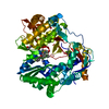
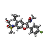
Mass: 446.492 Da / Num. of mol.: 1 / Source method: obtained synthetically / Formula: C22H23FN2O5S
#3: Chemical ChemComp-GOL / GLYCEROL / GLYCERIN / PROPANE-1,2,3-TRIOL / Glycerol


Mass: 92.094 Da / Num. of mol.: 2 / Source method: obtained synthetically / Formula: C3H8O3
#4: Water ChemComp-HOH / water / Water


Mass: 18.015 Da / Num. of mol.: 369 / Source method: isolated from a natural source / Formula: H2O

-
Experimental details

-
Experiment

ExperimentMethod: X-RAY DIFFRACTION / Number of used crystals: 1

-
Sample preparation

CrystalDensity Matthews: 2.2 Å3/Da / Density % sol: 44.19 %
Crystal growTemperature: 293 K / Method: vapor diffusion, sitting drop / pH: 4.9
Details: 50 mM NA CITRATE, 26% PEG 4000, 7.5% GLYCEROL, pH 4.9, VAPOR DIFFUSION, SITTING DROP, temperature 293K

-
Data collection

DiffractionMean temperature: 100 K
Diffraction sourceSource: SYNCHROTRON / Site: SSRL / Beamline: BL9-2 / Wavelength: 0.97946 Å
DetectorType: MARMOSAIC 325 mm CCD / Detector: CCD / Date: Nov 15, 2006
RadiationMonochromator: DOUBLE crystal si(111) / Protocol: SINGLE WAVELENGTH / Monochromatic (M) / Laue (L): M / Scattering type: x-ray
Radiation wavelengthWavelength: 0.97946 Å / Relative weight: 1
ReflectionResolution: 1.8→48.11 Å / Num. all: 53656 / Num. obs: 50278 / % possible obs: 93.7 % / Observed criterion σ(F): 0 / Observed criterion σ(I): 0 / Redundancy: 6.4 % / Biso Wilson estimate: 19 Å2 / Rsym value: 6.5 / Net I/σ(I): 27.5
Reflection shellResolution: 1.8→1.86 Å / Redundancy: 3 % / Mean I/σ(I) obs: 1.91 / Num. unique all: 3510 / Rsym value: 50.7 / % possible all: 66.7

-
Processing

Software
NameVersionClassification
Blu-Icedata collection
PHASERphasing
REFMAC5.2.0019refinement
HKL-2000data reduction
HKL-2000data scaling
RefinementMethod to determine structure: MOLECULAR REPLACEMENT
Starting model: pdb entry 2GIQ
Resolution: 1.8→40 Å / Cor.coef. Fo:Fc: 0.95 / Cor.coef. Fo:Fc free: 0.927 / SU B: 2.725 / SU ML: 0.086 / Cross valid method: THROUGHOUT / σ(F): 0 / σ(I): 0 / ESU R: 0.146 / ESU R Free: 0.133 / Stereochemistry target values: MAXIMUM LIKELIHOOD / Details: HYDROGENS HAVE BEEN ADDED IN THE RIDING POSITIONS
RfactorNum. reflection% reflectionSelection details
Rfree0.22337 2510 5 %RANDOM
Rwork0.1897 ---
all0.1914 53656 --
obs0.1914 47686 93.92 %-
Solvent computationIon probe radii: 0.8 Å / Shrinkage radii: 0.8 Å / VDW probe radii: 1.2 Å / Solvent model: MASK
Displacement parametersBiso mean: 16.376 Å2
Baniso -1Baniso -2Baniso -3
1--0.32 Å20 Å20 Å2
2--0.74 Å20 Å2
3----0.42 Å2
Refinement stepCycle: LAST / Resolution: 1.8→40 Å
ProteinNucleic acidLigandSolventTotal
Num. atoms4364 0 43 369 4776
Refine LS restraints
Refine-IDTypeDev idealDev ideal targetNumber
X-RAY DIFFRACTIONr_bond_refined_d0.0080.0224552
X-RAY DIFFRACTIONr_bond_other_d0.0010.023130
X-RAY DIFFRACTIONr_angle_refined_deg1.1571.9726186
X-RAY DIFFRACTIONr_angle_other_deg0.89737601
X-RAY DIFFRACTIONr_dihedral_angle_1_deg5.3565570
X-RAY DIFFRACTIONr_dihedral_angle_2_deg32.78622.432185
X-RAY DIFFRACTIONr_dihedral_angle_3_deg12.43415769
X-RAY DIFFRACTIONr_dihedral_angle_4_deg12.9741540
X-RAY DIFFRACTIONr_chiral_restr0.0650.2694
X-RAY DIFFRACTIONr_gen_planes_refined0.0040.025044
X-RAY DIFFRACTIONr_gen_planes_other0.0010.02943
X-RAY DIFFRACTIONr_nbd_refined0.2060.2964
X-RAY DIFFRACTIONr_nbd_other0.1870.23285
X-RAY DIFFRACTIONr_nbtor_refined0.1730.22244
X-RAY DIFFRACTIONr_nbtor_other0.0820.22329
X-RAY DIFFRACTIONr_xyhbond_nbd_refined0.1480.2314
X-RAY DIFFRACTIONr_xyhbond_nbd_other
X-RAY DIFFRACTIONr_metal_ion_refined
X-RAY DIFFRACTIONr_metal_ion_other
X-RAY DIFFRACTIONr_symmetry_vdw_refined0.1390.210
X-RAY DIFFRACTIONr_symmetry_vdw_other0.2130.263
X-RAY DIFFRACTIONr_symmetry_hbond_refined0.180.216
X-RAY DIFFRACTIONr_symmetry_hbond_other
X-RAY DIFFRACTIONr_symmetry_metal_ion_refined
X-RAY DIFFRACTIONr_symmetry_metal_ion_other
X-RAY DIFFRACTIONr_mcbond_it0.8171.53675
X-RAY DIFFRACTIONr_mcbond_other0.1141.51132
X-RAY DIFFRACTIONr_mcangle_it0.90724577
X-RAY DIFFRACTIONr_scbond_it1.57732045
X-RAY DIFFRACTIONr_scangle_it2.2794.51606
X-RAY DIFFRACTIONr_rigid_bond_restr
X-RAY DIFFRACTIONr_sphericity_free
X-RAY DIFFRACTIONr_sphericity_bonded
LS refinement shellResolution: 1.8→1.849 Å / Total num. of bins used: 20
RfactorNum. reflection% reflection
Rfree0.345 121 -
Rwork0.258 2302 -
obs-2423 61.81 %

+
About Yorodumi

-
News

-
Feb 9, 2022. New format data for meta-information of EMDB entries

New format data for meta-information of EMDB entries

  • Version 3 of the EMDB header file is now the official format.
  • The previous official version 1.9 will be removed from the archive.

Related info.:EMDB header

External links:wwPDB to switch to version 3 of the EMDB data model

-
Aug 12, 2020. Covid-19 info

Covid-19 info

URL: https://pdbj.org/emnavi/covid19.php

New page: Covid-19 featured information page in EM Navigator.

Related info.:Covid-19 info / Mar 5, 2020. Novel coronavirus structure data

+
Mar 5, 2020. Novel coronavirus structure data

Novel coronavirus structure data

Related info.:Yorodumi Speices / Aug 12, 2020. Covid-19 info

External links:COVID-19 featured content - PDBj / Molecule of the Month (242):Coronavirus Proteases

+
Jan 31, 2019. EMDB accession codes are about to change! (news from PDBe EMDB page)

EMDB accession codes are about to change! (news from PDBe EMDB page)

  • The allocation of 4 digits for EMDB accession codes will soon come to an end. Whilst these codes will remain in use, new EMDB accession codes will include an additional digit and will expand incrementally as the available range of codes is exhausted. The current 4-digit format prefixed with “EMD-” (i.e. EMD-XXXX) will advance to a 5-digit format (i.e. EMD-XXXXX), and so on. It is currently estimated that the 4-digit codes will be depleted around Spring 2019, at which point the 5-digit format will come into force.
  • The EM Navigator/Yorodumi systems omit the EMD- prefix.

Related info.:Q: What is EMD? / ID/Accession-code notation in Yorodumi/EM Navigator

External links:EMDB Accession Codes are Changing Soon! / Contact to PDBj

+
Jul 12, 2017. Major update of PDB

Major update of PDB

  • wwPDB released updated PDB data conforming to the new PDBx/mmCIF dictionary.
  • This is a major update changing the version number from 4 to 5, and with Remediation, in which all the entries are updated.
  • In this update, many items about electron microscopy experimental information are reorganized (e.g. em_software).
  • Now, EM Navigator and Yorodumi are based on the updated data.

External links:wwPDB Remediation / Enriched Model Files Conforming to OneDep Data Standards Now Available in the PDB FTP Archive

-
Yorodumi

Thousand views of thousand structures

  • Yorodumi is a browser for structure data from EMDB, PDB, SASBDB, etc.
  • This page is also the successor to EM Navigator detail page, and also detail information page/front-end page for Omokage search.
  • The word "yorodu" (or yorozu) is an old Japanese word meaning "ten thousand". "mi" (miru) is to see.

Related info.:EMDB / PDB / SASBDB / Comparison of 3 databanks / Yorodumi Search / Aug 31, 2016. New EM Navigator & Yorodumi / Yorodumi Papers / Jmol/JSmol / Function and homology information / Changes in new EM Navigator and Yorodumi

Read more