 Movie
Movie Controller


+ Open data

- Basic information
| Entry | Database: PDB / ID: 3fpw | ||||||
|---|---|---|---|---|---|---|---|
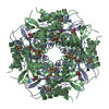
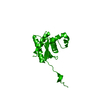
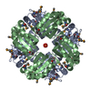
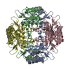
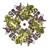
| Title | Crystal Structure of HbpS with bound iron | ||||||
 Components Components | Extracellular haem-binding protein | ||||||
 Keywords Keywords | HEME binding protein / haem binding | ||||||
| Function / homology |  Function and homology information Function and homology information: / Haem-degrading domain / Corrinoid adenosyltransferase PduO/GlcC-like / Corrinoid adenosyltransferase PduO/GlcC-like superfamily / Haem degrading protein HbpS-like / Beta-Lactamase / Twin arginine translocation (Tat) signal profile. / Twin-arginine translocation pathway, signal sequence / 2-Layer Sandwich / Alpha Beta Similarity search - Domain/homology | ||||||
| Biological species |  Streptomyces reticuli (bacteria) Streptomyces reticuli (bacteria) | ||||||
| Method |  X-RAY DIFFRACTION / SYNCHROTRON / MOLECULAR REPLACEMENT / Resolution: 1.6 Å X-RAY DIFFRACTION / SYNCHROTRON / MOLECULAR REPLACEMENT / Resolution: 1.6 Å | ||||||
 Authors Authors | Ortiz de Orue Lucana, D. / Bogel, G. / Zou, P. / Groves, M.R. | ||||||
 Citation Citation | Journal: J.Mol.Biol. / Year: 2009 Title: The oligomeric assembly of the novel haem-degrading protein HbpS is essential for interaction with its cognate two-component sensor kinase Authors: Ortiz de Orue Lucana, D. / Bogel, G. / Zou, P. / Groves, M.R. #1: Journal: Acta Crystallogr.,Sect.F / Year: 2008 Title: Crystallization and preliminary characterization of a novel haem-binding protein of Streptomyces reticuli Authors: Zou, P. / Groves, M.R. / Viale-Bouroncle, S.D. / Ortiz de Orue Lucana, D. | ||||||
| History |
|
- Structure visualization
| Structure viewer | Molecule: MolmilJmol/JSmol |
|---|
- Downloads & links
-Download
| PDBx/mmCIF format | 3fpw.cif.gz | 71.6 KB | Display | PDBx/mmCIF format |
|---|---|---|---|---|
| PDB format | pdb3fpw.ent.gz | 52.6 KB | Display | PDB format |
| PDBx/mmJSON format | 3fpw.json.gz | Tree view | PDBx/mmJSON format | |
| Others |  Other downloads Other downloads |
-Validation report
| Arichive directory | https://data.pdbj.org/pub/pdb/validation_reports/fp/3fpwftp://data.pdbj.org/pub/pdb/validation_reports/fp/3fpw | HTTPS FTP |
|---|
-Related structure data
| Related structure data | 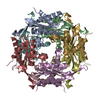 3fpvSC S: Starting model for refinement C: citing same article ( |
|---|---|
| Similar structure data |

-Links
 PDBj
PDBj

- Assembly
| Deposited unit | 
| ||||||||||||
|---|---|---|---|---|---|---|---|---|---|---|---|---|---|
| 1 | x 8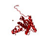
| ||||||||||||
| Unit cell |
| ||||||||||||
| Components on special symmetry positions |
| ||||||||||||
| Details | BIOPHYSICAL EXPERIMENTS INDICATE THAT THE IN VITRO SOLUTION STATE IS OCTOMERIC |
-Components
| #1: Protein | Mass: 18921.221 Da / Num. of mol.: 1 Source method: isolated from a genetically manipulated source Source: (gene. exp.) Streptomyces reticuli (bacteria) / Gene: hbpS / Plasmid: pETM11 / Production host: | ||||
|---|---|---|---|---|---|
| #2: Chemical |   Mass: 55.845 Da / Num. of mol.: 2 / Source method: obtained synthetically / Formula: Fe Mass: 55.845 Da / Num. of mol.: 2 / Source method: obtained synthetically / Formula: Fe#3: Chemical | ChemComp-PO4 /   Mass: 94.971 Da / Num. of mol.: 4 / Source method: obtained synthetically / Formula: PO4 Mass: 94.971 Da / Num. of mol.: 4 / Source method: obtained synthetically / Formula: PO4#4: Water | ChemComp-HOH / |  Mass: 18.015 Da / Num. of mol.: 133 / Source method: isolated from a natural source / Formula: H2O Mass: 18.015 Da / Num. of mol.: 133 / Source method: isolated from a natural source / Formula: H2O |
-Experimental details
-Experiment
| Experiment | Method: X-RAY DIFFRACTION / Number of used crystals: 1 |
|---|
- Sample preparation
| Crystal | Density Matthews: 1.605033 Å3/Da / Density % sol: 23.36606 % |
|---|---|
| Crystal grow | Temperature: 298 K / Method: vapor diffusion, hanging drop / pH: 5.8 Details: 40% w/v PEG 400, 5% w/v PEG 3000, 100mM MES pH 5.8; Protein concentration of 14 mg/ml and preincubated with haemin, VAPOR DIFFUSION, HANGING DROP, temperature 298K |
-Data collection
| Diffraction | Mean temperature: 100 K |
|---|---|
| Diffraction source | Source: SYNCHROTRON / Site: EMBL/DESY, HAMBURG  / Beamline: X12 / Wavelength: 1 Å / Beamline: X12 / Wavelength: 1 Å |
| Detector | Type: MARMOSAIC 225 mm CCD / Detector: CCD / Date: Nov 12, 2007 |
| Radiation | Monochromator: Silicon (111) / Protocol: SINGLE WAVELENGTH / Monochromatic (M) / Laue (L): M / Scattering type: x-ray |
| Radiation wavelength | Wavelength: 1 Å / Relative weight: 1 |
| Reflection | Resolution: 1.6→20 Å / Num. all: 16602 / Num. obs: 16582 / % possible obs: 99.9 % / Observed criterion σ(F): -3 / Observed criterion σ(I): -3 / Redundancy: 20.23 % / Rmerge(I) obs: 0.076 / Net I/σ(I): 28.73 |
| Reflection shell | Resolution: 1.6→1.7 Å / Redundancy: 85.17 % / Rmerge(I) obs: 0.721 / Mean I/σ(I) obs: 4.7 / Num. unique all: 2712 / % possible all: 100 |
- Processing
| Software |
| |||||||||||||||||||||||||||||||||
|---|---|---|---|---|---|---|---|---|---|---|---|---|---|---|---|---|---|---|---|---|---|---|---|---|---|---|---|---|---|---|---|---|---|---|
| Refinement | Method to determine structure: MOLECULAR REPLACEMENT Starting model: PDB ENTRY 3FPV Resolution: 1.6→18.8 Å / Cor.coef. Fo:Fc: 0.971 / Cor.coef. Fo:Fc free: 0.964 / SU B: 3.207 / SU ML: 0.051 / Cross valid method: THROUGHOUT / σ(F): -3 / σ(I): -3 / ESU R: 0.12 / ESU R Free: 0.084 / Stereochemistry target values: MAXIMUM LIKELIHOOD / Details: HYDROGENS HAVE BEEN ADDED IN THE RIDING POSITIONS
| |||||||||||||||||||||||||||||||||
| Solvent computation | Ion probe radii: 0.8 Å / Shrinkage radii: 0.8 Å / VDW probe radii: 1.2 Å / Solvent model: MASK | |||||||||||||||||||||||||||||||||
| Displacement parameters | Biso mean: 21.342 Å2
| |||||||||||||||||||||||||||||||||
| Refinement step | Cycle: LAST / Resolution: 1.6→18.8 Å
| |||||||||||||||||||||||||||||||||
| Refine LS restraints |
| |||||||||||||||||||||||||||||||||
| LS refinement shell | Resolution: 1.6→1.641 Å / Total num. of bins used: 20
|
