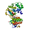
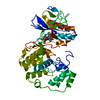
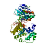
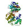
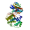
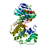
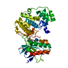
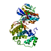
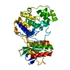
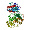
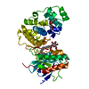
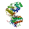
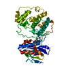
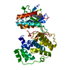
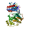
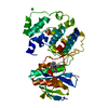
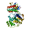
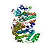
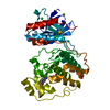
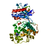
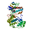
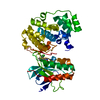
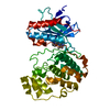
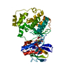
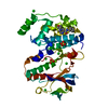
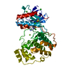
[English] 日本語
Yorodumi
- PDB-3fml: P38 kinase crystal structure in complex with RO6224 -

+
Open data


ID or keywords:

Loading...

-
Basic information

Entry
Database: PDB / ID: 3fml
TitleP38 kinase crystal structure in complex with RO6224
ComponentsMitogen-activated protein kinase 14MAPK14
KeywordsSIGNALING PROTEIN / TRANSFERASE / P38 / MAP KINASE / SERINE/THREONINE KINASE / ATP-binding / Kinase / Nucleotide-binding / Nucleus / Phosphoprotein / Serine/threonine-protein kinase
Function / homology
Function and homology information


positive regulation of cyclase activity / Activation of PPARGC1A (PGC-1alpha) by phosphorylation / CD163 mediating an anti-inflammatory response / regulation of synaptic membrane adhesion / stress-induced premature senescence / cell surface receptor protein serine/threonine kinase signaling pathway / 3'-UTR-mediated mRNA stabilization / KSRP (KHSRP) binds and destabilizes mRNA / DSCAM interactions / cartilage condensation ...positive regulation of cyclase activity / Activation of PPARGC1A (PGC-1alpha) by phosphorylation / CD163 mediating an anti-inflammatory response / regulation of synaptic membrane adhesion / stress-induced premature senescence / cell surface receptor protein serine/threonine kinase signaling pathway / 3'-UTR-mediated mRNA stabilization / KSRP (KHSRP) binds and destabilizes mRNA / DSCAM interactions / cartilage condensation / cellular response to UV-B / Platelet sensitization by LDL / stress-activated protein kinase signaling cascade / mitogen-activated protein kinase p38 binding / positive regulation of myoblast fusion / positive regulation of muscle cell differentiation / negative regulation of hippo signaling / positive regulation of myotube differentiation / NFAT protein binding / Myogenesis / glucose import / response to dietary excess / Activation of the AP-1 family of transcription factors / ERK/MAPK targets / regulation of cytokine production involved in inflammatory response / p38MAPK cascade / fatty acid oxidation / cellular response to lipoteichoic acid / MAP kinase kinase activity / response to muramyl dipeptide / RHO GTPases Activate NADPH Oxidases / regulation of ossification / MAP kinase activity / mitogen-activated protein kinase / cellular response to vascular endothelial growth factor stimulus / positive regulation of myoblast differentiation / chondrocyte differentiation / vascular endothelial growth factor receptor signaling pathway / stress-activated MAPK cascade / skeletal muscle tissue development / signal transduction in response to DNA damage / lipopolysaccharide-mediated signaling pathway / negative regulation of inflammatory response to antigenic stimulus / positive regulation of cardiac muscle cell proliferation / p38MAPK events / striated muscle cell differentiation / response to muscle stretch / positive regulation of brown fat cell differentiation / positive regulation of interleukin-12 production / osteoclast differentiation / positive regulation of erythrocyte differentiation / placenta development / DNA damage checkpoint signaling / activated TAK1 mediates p38 MAPK activation / cellular response to ionizing radiation / stem cell differentiation / positive regulation of glucose import / negative regulation of canonical Wnt signaling pathway / response to insulin / NOD1/2 Signaling Pathway / bone development / cell morphogenesis / platelet activation / osteoblast differentiation / cellular response to virus / VEGFA-VEGFR2 Pathway / spindle pole / positive regulation of protein import into nucleus / ADP signalling through P2Y purinoceptor 1 / glucose metabolic process / positive regulation of reactive oxygen species metabolic process / chemotaxis / cellular senescence / cellular response to tumor necrosis factor / protein phosphatase binding / peptidyl-serine phosphorylation / angiogenesis / Oxidative Stress Induced Senescence / secretory granule lumen / cellular response to lipopolysaccharide / Regulation of TP53 Activity through Phosphorylation / ficolin-1-rich granule lumen / transcription by RNA polymerase II / cell surface receptor signaling pathway / intracellular signal transduction / nuclear speck / protein serine kinase activity / protein serine/threonine kinase activity / glutamatergic synapse / apoptotic process / Neutrophil degranulation / positive regulation of gene expression / regulation of transcription by RNA polymerase II / enzyme binding / signal transduction / positive regulation of transcription by RNA polymerase II / mitochondrion / extracellular region / nucleoplasm / ATP binding
Similarity search - Function
Mitogen-activated protein kinase 14 / Mitogen-activated protein (MAP) kinase p38-like / Mitogen-activated protein (MAP) kinase, conserved site / MAP kinase signature. / Transferase(Phosphotransferase) domain 1 / Transferase(Phosphotransferase); domain 1 / Phosphorylase Kinase; domain 1 / Phosphorylase Kinase; domain 1 / Protein kinase domain / Serine/Threonine protein kinases, catalytic domain ...Mitogen-activated protein kinase 14 / Mitogen-activated protein (MAP) kinase p38-like / Mitogen-activated protein (MAP) kinase, conserved site / MAP kinase signature. / Transferase(Phosphotransferase) domain 1 / Transferase(Phosphotransferase); domain 1 / Phosphorylase Kinase; domain 1 / Phosphorylase Kinase; domain 1 / Protein kinase domain / Serine/Threonine protein kinases, catalytic domain / Protein kinase, ATP binding site / Protein kinases ATP-binding region signature. / Protein kinase domain profile. / Protein kinase domain / Protein kinase-like domain superfamily / 2-Layer Sandwich / Orthogonal Bundle / Mainly Alpha / Alpha Beta
Similarity search - Domain/homology
Chem-FML / Mitogen-activated protein kinase 14
Similarity search - Component
Biological speciesHomo sapiens (human)
MethodX-RAY DIFFRACTION / SYNCHROTRON / FOURIER SYNTHESIS / Resolution: 2.1 Å
AuthorsKuglstatter, A. / Ghate, M.
CitationJournal: To be Published
Title: P38 kinase crystal structure in complex with RO6224
Authors: Soth, M. / Kuglstatter, A. / Gabriel, T.
History
DepositionDec 22, 2008Deposition site: RCSB / Processing site: RCSB
Revision 1.0Dec 22, 2009Provider: repository / Type: Initial release
Revision 1.1Jul 13, 2011Group: Advisory / Refinement description / Version format compliance
Revision 1.2Jul 24, 2019Group: Data collection / Refinement description / Category: software
Item: _software.classification / _software.name / _software.version
Revision 1.3Sep 6, 2023Group: Data collection / Database references ...Data collection / Database references / Derived calculations / Refinement description
Category: chem_comp_atom / chem_comp_bond ...chem_comp_atom / chem_comp_bond / database_2 / pdbx_initial_refinement_model / struct_ref_seq_dif / struct_site
Item: _database_2.pdbx_DOI / _database_2.pdbx_database_accession ..._database_2.pdbx_DOI / _database_2.pdbx_database_accession / _struct_ref_seq_dif.details / _struct_site.pdbx_auth_asym_id / _struct_site.pdbx_auth_comp_id / _struct_site.pdbx_auth_seq_id

-
Structure visualization

Structure viewerMolecule:
MolmilJmol/JSmol

Downloads & links

-
Assembly

Deposited unit
A: Mitogen-activated protein kinase 14
hetero molecules


Theoretical massNumber of molelcules
Total (without water)43,1322
Polymers42,7491
Non-polymers3831
Water2,810156
1


  • Idetical with deposited unit
  • defined by author&software
TypeNameSymmetry operationNumber
identity operation1_555x,y,z1
MethodPISA
Unit cell
Length a, b, c (Å)45.272, 86.110, 124.963
Angle α, β, γ (deg.)90.00, 90.00, 90.00
Int Tables number19
Space group name H-MP212121
DetailsThe asymmetric unit of the crystal structure contains one biological unit.

-
Components

#1: Protein Mitogen-activated protein kinase 14 / MAPK14 / Mitogen-activated protein kinase p38 alpha / MAP kinase p38 alpha / Cytokine suppressive anti- ...Mitogen-activated protein kinase p38 alpha / MAP kinase p38 alpha / Cytokine suppressive anti-inflammatory drug-binding protein / CSAID-binding protein / CSBP / MAX-interacting protein 2 / MAP kinase MXI2 / SAPK2A


Mass: 42748.715 Da / Num. of mol.: 1
Source method: isolated from a genetically manipulated source
Source: (gene. exp.) Homo sapiens (human) / Gene: CSBP, CSBP1, CSBP2, CSPB1, MAPK14, MXI2 / Production host: ESCHERICHIA COLI (E. coli)
References: UniProt: Q16539, mitogen-activated protein kinase
#2: Chemical ChemComp-FML / 6-(2,4-difluorophenoxy)-N-[(1S)-1-methyl-2-(methylsulfonyl)ethyl]-1H-pyrazolo[3,4-d]pyrimidin-3-amine


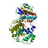
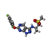
Mass: 383.373 Da / Num. of mol.: 1 / Source method: obtained synthetically / Formula: C15H15F2N5O3S
#3: Water ChemComp-HOH / water / Water


Mass: 18.015 Da / Num. of mol.: 156 / Source method: isolated from a natural source / Formula: H2O

-
Experimental details

-
Experiment

ExperimentMethod: X-RAY DIFFRACTION / Number of used crystals: 1

-
Sample preparation

CrystalDensity Matthews: 2.85 Å3/Da / Density % sol: 56.83 %
Crystal growTemperature: 290 K / Method: vapor diffusion, hanging drop / pH: 7.6
Details: 50 mM Hepes pH 7.6, 50 mM CaCl2, 17% PEG 3350, VAPOR DIFFUSION, HANGING DROP, temperature 290K

-
Data collection

DiffractionMean temperature: 100 K
Diffraction sourceSource: SYNCHROTRON / Site: ALS / Beamline: 5.0.1 / Wavelength: 1 Å
DetectorType: ADSC QUANTUM 210 / Detector: CCD
RadiationProtocol: SINGLE WAVELENGTH / Monochromatic (M) / Laue (L): M / Scattering type: x-ray
Radiation wavelengthWavelength: 1 Å / Relative weight: 1
ReflectionResolution: 2.1→50 Å / Num. obs: 25944 / % possible obs: 88.5 % / Redundancy: 4.1 % / Biso Wilson estimate: 41.9 Å2 / Rsym value: 0.062
Reflection shellResolution: 2.1→2.18 Å / Redundancy: 3 % / Mean I/σ(I) obs: 3.3 / Num. unique all: 2407 / Rsym value: 0.354 / % possible all: 83.6

-
Processing

Software
NameVersionClassification
REFMAC5.2.0005refinement
CNSrefinement
ADSCQuantumdata collection
DENZOdata reduction
SCALEPACKdata scaling
CNSphasing
RefinementMethod to determine structure: FOURIER SYNTHESIS
Starting model: PDB entry 1P38

1p38
PDB Unreleased entry


Resolution: 2.1→33.73 Å / Cor.coef. Fo:Fc: 0.947 / Cor.coef. Fo:Fc free: 0.94 / SU B: 10.359 / SU ML: 0.126 / TLS residual ADP flag: LIKELY RESIDUAL / Cross valid method: THROUGHOUT / ESU R: 0.23 / ESU R Free: 0.19 / Stereochemistry target values: MAXIMUM LIKELIHOOD
RfactorNum. reflection% reflectionSelection details
Rfree0.24518 1291 5 %RANDOM
Rwork0.21444 ---
obs0.21599 24598 88.27 %-
Solvent computationIon probe radii: 0.8 Å / Shrinkage radii: 0.8 Å / VDW probe radii: 1.2 Å / Solvent model: BABINET MODEL WITH MASK
Displacement parametersBiso mean: 53.223 Å2
Baniso -1Baniso -2Baniso -3
1--1.45 Å20 Å20 Å2
2---0.28 Å20 Å2
3---1.73 Å2
Refinement stepCycle: LAST / Resolution: 2.1→33.73 Å
ProteinNucleic acidLigandSolventTotal
Num. atoms2778 0 26 156 2960
Refine LS restraints
Refine-IDTypeDev idealDev ideal targetNumber
X-RAY DIFFRACTIONr_bond_refined_d0.0070.0222872
X-RAY DIFFRACTIONr_bond_other_d
X-RAY DIFFRACTIONr_angle_refined_deg1.0241.9763903
X-RAY DIFFRACTIONr_angle_other_deg
X-RAY DIFFRACTIONr_dihedral_angle_1_deg4.2345339
X-RAY DIFFRACTIONr_dihedral_angle_2_deg36.05323.971136
X-RAY DIFFRACTIONr_dihedral_angle_3_deg11.62115500
X-RAY DIFFRACTIONr_dihedral_angle_4_deg15.781519
X-RAY DIFFRACTIONr_chiral_restr0.0640.2438
X-RAY DIFFRACTIONr_gen_planes_refined0.0030.022168
X-RAY DIFFRACTIONr_gen_planes_other
X-RAY DIFFRACTIONr_nbd_refined0.2080.21303
X-RAY DIFFRACTIONr_nbd_other
X-RAY DIFFRACTIONr_nbtor_refined0.3030.21984
X-RAY DIFFRACTIONr_nbtor_other
X-RAY DIFFRACTIONr_xyhbond_nbd_refined0.1270.2173
X-RAY DIFFRACTIONr_xyhbond_nbd_other
X-RAY DIFFRACTIONr_metal_ion_refined
X-RAY DIFFRACTIONr_metal_ion_other
X-RAY DIFFRACTIONr_symmetry_vdw_refined0.1710.243
X-RAY DIFFRACTIONr_symmetry_vdw_other
X-RAY DIFFRACTIONr_symmetry_hbond_refined0.1110.28
X-RAY DIFFRACTIONr_symmetry_hbond_other
X-RAY DIFFRACTIONr_symmetry_metal_ion_refined
X-RAY DIFFRACTIONr_symmetry_metal_ion_other
X-RAY DIFFRACTIONr_mcbond_it0.7711.51782
X-RAY DIFFRACTIONr_mcbond_other
X-RAY DIFFRACTIONr_mcangle_it1.13522790
X-RAY DIFFRACTIONr_scbond_it1.37831265
X-RAY DIFFRACTIONr_scangle_it2.084.51113
X-RAY DIFFRACTIONr_rigid_bond_restr
X-RAY DIFFRACTIONr_sphericity_free
X-RAY DIFFRACTIONr_sphericity_bonded
LS refinement shellResolution: 2.1→2.155 Å / Total num. of bins used: 20
RfactorNum. reflection% reflection
Rfree0.296 98 -
Rwork0.261 1552 -
obs--76.28 %
Refinement TLS params.Method: refined / Origin x: 21.3503 Å / Origin y: 17.3845 Å / Origin z: 21.6989 Å
111213212223313233
T-0.004 Å20.016 Å2-0.0125 Å2--0.0056 Å20.0145 Å2---0.012 Å2
L0.1984 °20.1589 °2-0.1786 °2-0.2459 °2-0.0193 °2--0.2904 °2
S-0.013 Å °0.0176 Å °-0.0454 Å °-0.008 Å °0.0228 Å °-0.0164 Å °-0.0117 Å °-0.0249 Å °-0.0098 Å °
Refinement TLS group
IDRefine-IDRefine TLS-IDAuth asym-IDAuth seq-ID
1X-RAY DIFFRACTION1A1001 - 2152
2X-RAY DIFFRACTION1A361
3X-RAY DIFFRACTION1A4 - 352

+
About Yorodumi

-
News

-
Feb 9, 2022. New format data for meta-information of EMDB entries

New format data for meta-information of EMDB entries

  • Version 3 of the EMDB header file is now the official format.
  • The previous official version 1.9 will be removed from the archive.

Related info.:EMDB header

External links:wwPDB to switch to version 3 of the EMDB data model

-
Aug 12, 2020. Covid-19 info

Covid-19 info

URL: https://pdbj.org/emnavi/covid19.php

New page: Covid-19 featured information page in EM Navigator.

Related info.:Covid-19 info / Mar 5, 2020. Novel coronavirus structure data

+
Mar 5, 2020. Novel coronavirus structure data

Novel coronavirus structure data

Related info.:Yorodumi Speices / Aug 12, 2020. Covid-19 info

External links:COVID-19 featured content - PDBj / Molecule of the Month (242):Coronavirus Proteases

+
Jan 31, 2019. EMDB accession codes are about to change! (news from PDBe EMDB page)

EMDB accession codes are about to change! (news from PDBe EMDB page)

  • The allocation of 4 digits for EMDB accession codes will soon come to an end. Whilst these codes will remain in use, new EMDB accession codes will include an additional digit and will expand incrementally as the available range of codes is exhausted. The current 4-digit format prefixed with “EMD-” (i.e. EMD-XXXX) will advance to a 5-digit format (i.e. EMD-XXXXX), and so on. It is currently estimated that the 4-digit codes will be depleted around Spring 2019, at which point the 5-digit format will come into force.
  • The EM Navigator/Yorodumi systems omit the EMD- prefix.

Related info.:Q: What is EMD? / ID/Accession-code notation in Yorodumi/EM Navigator

External links:EMDB Accession Codes are Changing Soon! / Contact to PDBj

+
Jul 12, 2017. Major update of PDB

Major update of PDB

  • wwPDB released updated PDB data conforming to the new PDBx/mmCIF dictionary.
  • This is a major update changing the version number from 4 to 5, and with Remediation, in which all the entries are updated.
  • In this update, many items about electron microscopy experimental information are reorganized (e.g. em_software).
  • Now, EM Navigator and Yorodumi are based on the updated data.

External links:wwPDB Remediation / Enriched Model Files Conforming to OneDep Data Standards Now Available in the PDB FTP Archive

-
Yorodumi

Thousand views of thousand structures

  • Yorodumi is a browser for structure data from EMDB, PDB, SASBDB, etc.
  • This page is also the successor to EM Navigator detail page, and also detail information page/front-end page for Omokage search.
  • The word "yorodu" (or yorozu) is an old Japanese word meaning "ten thousand". "mi" (miru) is to see.

Related info.:EMDB / PDB / SASBDB / Comparison of 3 databanks / Yorodumi Search / Aug 31, 2016. New EM Navigator & Yorodumi / Yorodumi Papers / Jmol/JSmol / Function and homology information / Changes in new EM Navigator and Yorodumi

Read more