- PDB-3fmc: CRYSTAL STRUCTURE OF a putative succinylglutamate desuccinylase /... -
+
Open data
ID or keywords:
Loading...
-
Basic information
Entry
Database: PDB / ID: 3fmc
Title
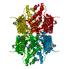
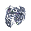
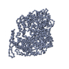
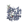
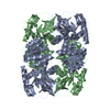
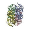
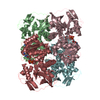
CRYSTAL STRUCTURE OF a putative succinylglutamate desuccinylase / aspartoacylase family protein (SAMA_0604) FROM SHEWANELLA AMAZONENSIS SB2B AT 1.80 A RESOLUTION
Mass: 18.015 Da / Num. of mol.: 1759 / Source method: isolated from a natural source / Formula: H2O
Has protein modification
Y
Sequence details
THE CONSTRUCT WAS EXPRESSED WITH A PURIFICATION TAG MGSDKIHHHHHHENLYFQG. THE TAG WAS REMOVED WITH ...THE CONSTRUCT WAS EXPRESSED WITH A PURIFICATION TAG MGSDKIHHHHHHENLYFQG. THE TAG WAS REMOVED WITH TEV PROTEASE LEAVING ONLY A GLYCINE (0) FOLLOWED BY THE TARGET SEQUENCE.
-
Experimental details
-
Experiment
Experiment
Method: X-RAY DIFFRACTION / Number of used crystals: 1
-
Sample preparation
Crystal
Density Matthews: 3.42 Å3/Da / Density % sol: 63.99 %
Crystal grow
Temperature: 293 K / Method: vapor diffusion, sitting drop / pH: 7.5 Details: NANODROP, 1.5M Li2SO4, 0.1M HEPES pH 7.5, VAPOR DIFFUSION, SITTING DROP, temperature 293K
Resolution: 1.8→29.735 Å / Num. obs: 184530 / % possible obs: 88.4 % / Redundancy: 4.8 % / Biso Wilson estimate: 18.527 Å2 / Rmerge(I) obs: 0.105 / Rsym value: 0.105 / Net I/σ(I): 5.074
Reflection shell
Diffraction-ID: 1
Resolution (Å)
Redundancy (%)
Rmerge(I) obs
Mean I/σ(I) obs
Num. measured all
Num. unique all
Rsym value
% possible all
1.8-1.85
3.6
0.572
1.3
38289
10674
0.572
69.7
1.85-1.9
3.6
0.43
1.8
37572
10381
0.43
69.3
1.9-1.95
3.7
0.362
2.1
37228
10127
0.362
69.8
1.95-2.01
3.9
0.338
2.2
53197
13532
0.338
95.8
2.01-2.08
4
0.274
2.7
51754
13089
0.274
95.5
2.08-2.15
4
0.233
3.2
50057
12619
0.233
95.3
2.15-2.23
4
0.205
3.5
48194
12130
0.205
94.9
2.23-2.32
4
0.179
4
46512
11671
0.179
94.9
2.32-2.43
4
0.158
4.4
44683
11153
0.158
94.4
2.43-2.55
4
0.139
5
42874
10649
0.139
94.1
2.55-2.68
5
0.146
4.7
50153
10095
0.146
93.7
2.68-2.85
5.4
0.134
5.1
51763
9530
0.134
93.3
2.85-3.04
6.2
0.117
5.6
55721
8937
0.117
92.9
3.04-3.29
6.8
0.1
6.3
55873
8275
0.1
92.4
3.29-3.6
6.8
0.084
7.4
51582
7593
0.084
91.9
3.6-4.02
6.8
0.077
7.7
46549
6851
0.077
91.3
4.02-4.65
6.8
0.072
8.2
40910
6030
0.072
90.6
4.65-5.69
6.8
0.071
8.2
34677
5078
0.071
89.9
5.69-8.05
6.9
0.073
8
26998
3934
0.073
88.8
8.05-29.735
6.7
0.066
7.2
14527
2182
0.066
85.5
-
Phasing
Phasing
Method: MAD
-
Processing
Software
Name
Version
Classification
NB
REFMAC
5.2.0019
refinement
PHENIX
refinement
SHELX
phasing
MolProbity
3beta29
modelbuilding
SCALA
3.2.5
datascaling
PDB_EXTRACT
3.006
dataextraction
MAR345
CCD
datacollection
MOSFLM
datareduction
SHELXD
phasing
autoSHARP
phasing
Refinement
Method to determine structure: MAD / Resolution: 1.8→29.735 Å / Cor.coef. Fo:Fc: 0.966 / Cor.coef. Fo:Fc free: 0.954 / Occupancy max: 1 / Occupancy min: 0.25 / SU B: 1.98 / SU ML: 0.062 / Cross valid method: THROUGHOUT / σ(F): 0 / ESU R: 0.1 / ESU R Free: 0.1 Stereochemistry target values: MAXIMUM LIKELIHOOD WITH PHASES Details: 1. HYDROGENS HAVE BEEN ADDED IN THE RIDING POSITIONS. 2. A MET-INHIBITION PROTOCOL WAS USED FOR SELENOMETHIONINE INCORPORATION DURING PROTEIN EXPRESSION. THE OCCUPANCY OF THE SE ATOMS IN THE ...Details: 1. HYDROGENS HAVE BEEN ADDED IN THE RIDING POSITIONS. 2. A MET-INHIBITION PROTOCOL WAS USED FOR SELENOMETHIONINE INCORPORATION DURING PROTEIN EXPRESSION. THE OCCUPANCY OF THE SE ATOMS IN THE MSE RESIDUES WAS REDUCED TO 0.75 FOR THE REDUCED SCATTERING POWER DUE TO PARTIAL S-MET INCORPORATION. 3. SULFATE (SO4) IONS FROM CRYSTALLIZATION CONDITION AND GLYCEROL (GOL) MOLECULES FROM CRYO SOLUTION ARE MODELED. 4. RAMACHANDRAN OUTLIER RESIDUES 174 IN ALL FOUR CHAINS ARE SUPPORTED BY ELECTRON DENSITY.
Rfactor
Num. reflection
% reflection
Selection details
Rfree
0.187
9270
5 %
RANDOM
Rwork
0.155
-
-
-
obs
0.157
184450
88.15 %
-
Solvent computation
Ion probe radii: 0.8 Å / Shrinkage radii: 0.8 Å / VDW probe radii: 1.2 Å / Solvent model: MASK
In the structure databanks used in Yorodumi, some data are registered as the other names, "COVID-19 virus" and "2019-nCoV". Here are the details of the virus and the list of structure data.
Jan 31, 2019. EMDB accession codes are about to change! (news from PDBe EMDB page)
EMDB accession codes are about to change! (news from PDBe EMDB page)
The allocation of 4 digits for EMDB accession codes will soon come to an end. Whilst these codes will remain in use, new EMDB accession codes will include an additional digit and will expand incrementally as the available range of codes is exhausted. The current 4-digit format prefixed with “EMD-” (i.e. EMD-XXXX) will advance to a 5-digit format (i.e. EMD-XXXXX), and so on. It is currently estimated that the 4-digit codes will be depleted around Spring 2019, at which point the 5-digit format will come into force.
The EM Navigator/Yorodumi systems omit the EMD- prefix.
Related info.:Q: What is EMD? / ID/Accession-code notation in Yorodumi/EM Navigator
Yorodumi is a browser for structure data from EMDB, PDB, SASBDB, etc.
This page is also the successor to EM Navigator detail page, and also detail information page/front-end page for Omokage search.
The word "yorodu" (or yorozu) is an old Japanese word meaning "ten thousand". "mi" (miru) is to see.
Related info.:EMDB / PDB / SASBDB / Comparison of 3 databanks / Yorodumi Search / Aug 31, 2016. New EM Navigator & Yorodumi / Yorodumi Papers / Jmol/JSmol / Function and homology information / Changes in new EM Navigator and Yorodumi
 Movie
Movie Controller

 Yorodumi
 Open data
 Basic information
 Components
 Keywords
 Function and homology information
 Shewanella amazonensis (bacteria)
 X-RAY DIFFRACTION /
  Authors
 Citation
 Structure visualization
 Downloads & links
 Other downloads
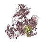
 PDBj
PDBj Assembly

 Mass: 96.063 Da / Num. of mol.: 5 / Source method: obtained synthetically / Formula: SO4
 Mass: 92.094 Da / Num. of mol.: 8 / Source method: obtained synthetically / Formula: C3H8O3
 Mass: 18.015 Da / Num. of mol.: 1759 / Source method: isolated from a natural source / Formula: H2O
 Sample preparation
 / Beamline: 23-ID-B / Wavelength: 0.94645, 0.97967
 Processing