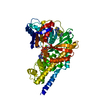
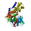
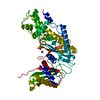
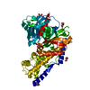
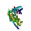
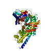
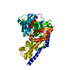
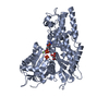
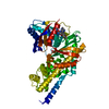
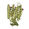
Defective GCK causes maturity-onset diabetes of the young 2 (MODY2) / glucose sensor activity / mannokinase activity / regulation of potassium ion transport / hexokinase / glucose catabolic process / fructokinase activity / glucokinase activity / glucose 6-phosphate metabolic process / NADP+ metabolic process ...Defective GCK causes maturity-onset diabetes of the young 2 (MODY2) / glucose sensor activity / mannokinase activity / regulation of potassium ion transport / hexokinase / glucose catabolic process / fructokinase activity / glucokinase activity / glucose 6-phosphate metabolic process / NADP+ metabolic process / Regulation of Glucokinase by Glucokinase Regulatory Protein / Defective TPR may confer susceptibility towards thyroid papillary carcinoma (TPC) / calcium ion import / D-glucose binding / cellular response to leptin stimulus / Glycolysis / canonical glycolysis / regulation of glycolytic process / Regulation of gene expression in beta cells / regulation of insulin secretion / negative regulation of gluconeogenesis / positive regulation of glycogen biosynthetic process / FOXO-mediated transcription of oxidative stress, metabolic and neuronal genes / response to glucose / intracellular glucose homeostasis / glycolytic process / glucose metabolic process / cellular response to insulin stimulus / positive regulation of insulin secretion / glucose homeostasis / mitochondrion / nucleoplasm / ATP binding / cytosol Similarity search - Function
In the structure databanks used in Yorodumi, some data are registered as the other names, "COVID-19 virus" and "2019-nCoV". Here are the details of the virus and the list of structure data.
Jan 31, 2019. EMDB accession codes are about to change! (news from PDBe EMDB page)
EMDB accession codes are about to change! (news from PDBe EMDB page)
The allocation of 4 digits for EMDB accession codes will soon come to an end. Whilst these codes will remain in use, new EMDB accession codes will include an additional digit and will expand incrementally as the available range of codes is exhausted. The current 4-digit format prefixed with “EMD-” (i.e. EMD-XXXX) will advance to a 5-digit format (i.e. EMD-XXXXX), and so on. It is currently estimated that the 4-digit codes will be depleted around Spring 2019, at which point the 5-digit format will come into force.
The EM Navigator/Yorodumi systems omit the EMD- prefix.
Related info.:Q: What is EMD? / ID/Accession-code notation in Yorodumi/EM Navigator
Yorodumi is a browser for structure data from EMDB, PDB, SASBDB, etc.
This page is also the successor to EM Navigator detail page, and also detail information page/front-end page for Omokage search.
The word "yorodu" (or yorozu) is an old Japanese word meaning "ten thousand". "mi" (miru) is to see.
Related info.:EMDB / PDB / SASBDB / Comparison of 3 databanks / Yorodumi Search / Aug 31, 2016. New EM Navigator & Yorodumi / Yorodumi Papers / Jmol/JSmol / Function and homology information / Changes in new EM Navigator and Yorodumi
 Movie
Movie Controller


 Open data
 Basic information
 Components
 Keywords
 Function and homology information
 Homo sapiens (human)
 X-RAY DIFFRACTION /
  Authors
 Citation
 Structure visualization
 Downloads & links
 Other downloads


 PDBj
PDBj


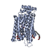
 Assembly
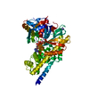


 Type: D-saccharide, beta linking / Mass: 180.156 Da / Num. of mol.: 1


 Mass: 506.196 Da / Num. of mol.: 1 / Source method: obtained synthetically / Formula: C10H17N6O12P3 / Comment: AMP-PNP, energy-carrying molecule analogue*YM
 Mass: 24.305 Da / Num. of mol.: 1 / Source method: obtained synthetically / Formula: Mg
 Mass: 39.098 Da / Num. of mol.: 1 / Source method: obtained synthetically / Formula: K
 Sample preparation
 / Beamline: X06SA / Wavelength: 1.0001 Å
 Processing