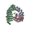
Inhibition of replication initiation of damaged DNA by RB1/E2F1 / ERKs are inactivated / Initiation of Nuclear Envelope (NE) Reformation / Cyclin A/B1/B2 associated events during G2/M transition / Co-inhibition by CTLA4 / mitotic sister chromatid cohesion, centromeric / Beta-catenin phosphorylation cascade / Co-stimulation by CD28 / Disassembly of the destruction complex and recruitment of AXIN to the membrane / Negative regulation of MAPK pathway ...Inhibition of replication initiation of damaged DNA by RB1/E2F1 / ERKs are inactivated / Initiation of Nuclear Envelope (NE) Reformation / Cyclin A/B1/B2 associated events during G2/M transition / Co-inhibition by CTLA4 / mitotic sister chromatid cohesion, centromeric / Beta-catenin phosphorylation cascade / Co-stimulation by CD28 / Disassembly of the destruction complex and recruitment of AXIN to the membrane / Negative regulation of MAPK pathway / Spry regulation of FGF signaling / Regulation of TP53 Degradation / Nonsense Mediated Decay (NMD) enhanced by the Exon Junction Complex (EJC) / centriole-centriole cohesion / Amplification of signal from unattached kinetochores via a MAD2 inhibitory signal / Cyclin D associated events in G1 / RAF activation / meiotic spindle elongation / Integration of energy metabolism / PP2A-mediated dephosphorylation of key metabolic factors / Mitotic Prometaphase / EML4 and NUDC in mitotic spindle formation / Degradation of beta-catenin by the destruction complex / Resolution of Sister Chromatid Cohesion / PKR-mediated signaling / meiotic chromosome segregation / mitotic sister chromatid separation / MASTL Facilitates Mitotic Progression / Turbulent (oscillatory, disturbed) flow shear stress activates signaling by PIEZO1 and integrins in endothelial cells / DARPP-32 events / protein phosphatase type 2A complex / RNA polymerase II CTD heptapeptide repeat S2 phosphatase activity / RNA polymerase II CTD heptapeptide repeat S7 phosphatase activity / protein serine/threonine phosphatase complex / regulation of meiotic cell cycle process involved in oocyte maturation / peptidyl-threonine dephosphorylation / attachment of spindle microtubules to kinetochore / meiotic sister chromatid cohesion, centromeric / INTAC complex / RHO GTPases Activate Formins / Separation of Sister Chromatids / Platelet sensitization by LDL / FAR/SIN/STRIPAK complex / Loss of Nlp from mitotic centrosomes / Recruitment of mitotic centrosome proteins and complexes / Loss of proteins required for interphase microtubule organization from the centrosome / ERK/MAPK targets / Recruitment of NuMA to mitotic centrosomes / Anchoring of the basal body to the plasma membrane / AURKA Activation by TPX2 / RNA polymerase II CTD heptapeptide repeat S5 phosphatase activity / PI5P, PP2A and IER3 Regulate PI3K/AKT Signaling / Regulation of glycolysis by fructose 2,6-bisphosphate metabolism / Inhibition of replication initiation of damaged DNA by RB1/E2F1 / female meiotic nuclear division / meiotic sister chromatid cohesion / Regulation of PLK1 Activity at G2/M Transition / protein phosphatase regulator activity / GABA receptor binding / condensed chromosome, centromeric region / APC truncation mutants have impaired AXIN binding / AXIN missense mutants destabilize the destruction complex / Truncations of AMER1 destabilize the destruction complex / protein antigen binding / ERKs are inactivated / Initiation of Nuclear Envelope (NE) Reformation / positive regulation of extrinsic apoptotic signaling pathway in absence of ligand / Beta-catenin phosphorylation cascade / Signaling by GSK3beta mutants / CTNNB1 S33 mutants aren't phosphorylated / CTNNB1 S37 mutants aren't phosphorylated / CTNNB1 S45 mutants aren't phosphorylated / CTNNB1 T41 mutants aren't phosphorylated / RNA polymerase II transcription initiation surveillance / Co-stimulation by CD28 / regulation of growth / Disassembly of the destruction complex and recruitment of AXIN to the membrane / negative regulation of epithelial to mesenchymal transition / Co-inhibition by CTLA4 / Platelet sensitization by LDL / protein-serine/threonine phosphatase / negative regulation of glycolytic process through fructose-6-phosphate / positive regulation of NLRP3 inflammasome complex assembly / ERK/MAPK targets / protein serine/threonine phosphatase activity / mesoderm development / vascular endothelial cell response to oscillatory fluid shear stress / T cell homeostasis / regulation of G1/S transition of mitotic cell cycle / regulation of cell differentiation / regulation of microtubule polymerization / phosphoprotein phosphatase activity / protein phosphatase activator activity / lateral plasma membrane / chromosome, centromeric region / DARPP-32 events / intrinsic apoptotic signaling pathway in response to DNA damage by p53 class mediator / negative regulation of hippo signaling / Cyclin A/B1/B2 associated events during G2/M transition / Nonsense Mediated Decay (NMD) enhanced by the Exon Junction Complex (EJC) Similarity search - Function
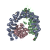
Shugoshin, C-terminal / Shugoshin, N-terminal coiled-coil domain / Shugoshin / Shugoshin C terminus / Shugoshin N-terminal coiled-coil region / Single helix bin / Protein phosphatase 2A, regulatory B subunit, B56 / Protein phosphatase 2A regulatory B subunit (B56 family) / : / : ...Shugoshin, C-terminal / Shugoshin, N-terminal coiled-coil domain / Shugoshin / Shugoshin C terminus / Shugoshin N-terminal coiled-coil region / Single helix bin / Protein phosphatase 2A, regulatory B subunit, B56 / Protein phosphatase 2A regulatory B subunit (B56 family) / : / : / HEAT repeat / HEAT repeat / : / PPP2R1A-like HEAT repeat / Serine/threonine specific protein phosphatases signature. / Protein phosphatase 2A homologues, catalytic domain. / Serine/threonine-specific protein phosphatase/bis(5-nucleosyl)-tetraphosphatase / HEAT repeat profile. / HEAT, type 2 / HEAT repeats / Metallo-dependent phosphatases / Purple Acid Phosphatase; chain A, domain 2 / Calcineurin-like phosphoesterase domain, ApaH type / Calcineurin-like phosphoesterase / Leucine-rich Repeat Variant / Metallo-dependent phosphatase-like / Leucine-rich Repeat Variant / Single alpha-helices involved in coiled-coils or other helix-helix interfaces / 4-Layer Sandwich / Armadillo-like helical / Alpha Horseshoe / Armadillo-type fold / Up-down Bundle / Mainly Alpha / Alpha Beta Similarity search - Domain/homology
In the structure databanks used in Yorodumi, some data are registered as the other names, "COVID-19 virus" and "2019-nCoV". Here are the details of the virus and the list of structure data.
Jan 31, 2019. EMDB accession codes are about to change! (news from PDBe EMDB page)
EMDB accession codes are about to change! (news from PDBe EMDB page)
The allocation of 4 digits for EMDB accession codes will soon come to an end. Whilst these codes will remain in use, new EMDB accession codes will include an additional digit and will expand incrementally as the available range of codes is exhausted. The current 4-digit format prefixed with “EMD-” (i.e. EMD-XXXX) will advance to a 5-digit format (i.e. EMD-XXXXX), and so on. It is currently estimated that the 4-digit codes will be depleted around Spring 2019, at which point the 5-digit format will come into force.
The EM Navigator/Yorodumi systems omit the EMD- prefix.
Related info.:Q: What is EMD? / ID/Accession-code notation in Yorodumi/EM Navigator
Yorodumi is a browser for structure data from EMDB, PDB, SASBDB, etc.
This page is also the successor to EM Navigator detail page, and also detail information page/front-end page for Omokage search.
The word "yorodu" (or yorozu) is an old Japanese word meaning "ten thousand". "mi" (miru) is to see.
Related info.:EMDB / PDB / SASBDB / Comparison of 3 databanks / Yorodumi Search / Aug 31, 2016. New EM Navigator & Yorodumi / Yorodumi Papers / Jmol/JSmol / Function and homology information / Changes in new EM Navigator and Yorodumi
 Movie
Movie Controller


 Open data
 Basic information
 Components
 Keywords
 Function and homology information
 Homo sapiens (human)
 Microcystis aeruginosa (bacteria)
 X-RAY DIFFRACTION /
  Authors
 Citation
 Structure visualization
 Downloads & links
 Other downloads


 PDBj
PDBj


 Assembly
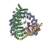
 Trichoplusia ni (cabbage looper)
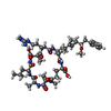
 Type: Oligopeptide / Class: Toxin / Mass: 1014.195 Da / Num. of mol.: 1 / Source method: obtained synthetically / Source: (synth.)

 Mass: 54.938 Da / Num. of mol.: 2 / Source method: obtained synthetically / Formula: Mn
 Sample preparation
 / Beamline: 5.0.2 / Wavelength: 1 Å
 Processing