 Movie
Movie Controller


+ Open data

- Basic information
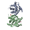
| Entry | Database: PDB / ID: 3fdq | ||||||
|---|---|---|---|---|---|---|---|
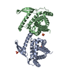
| Title | Recognition of AT-rich DNA binding sites by the MogR Repressor | ||||||
 Components Components |
| ||||||
 Keywords Keywords | DNA BINDING PROTEIN/DNA / Protein-DNA complex / Helix-turn-helix / minor groove binding / Cytoplasm / DNA-binding / Repressor / Transcription / Transcription regulation / Virulence / DNA BINDING PROTEIN-DNA COMPLEX | ||||||
| Function / homology |  Function and homology information Function and homology information: / DNA-binding transcription repressor activity / protein-DNA complex / transcription cis-regulatory region binding / negative regulation of DNA-templated transcription / DNA binding / cytoplasm Similarity search - Function | ||||||
| Biological species |  Listeria monocytogenes (bacteria) Listeria monocytogenes (bacteria) | ||||||
| Method |  X-RAY DIFFRACTION / SYNCHROTRON / SAD / Resolution: 1.75 Å X-RAY DIFFRACTION / SYNCHROTRON / SAD / Resolution: 1.75 Å | ||||||
 Authors Authors | Shen, A. / Higgins, D.E. / Panne, D. | ||||||
 Citation Citation | Journal: Structure / Year: 2009 Title: Recognition of AT-Rich DNA Binding Sites by the MogR Repressor. Authors: Shen, A. / Higgins, D.E. / Panne, D. | ||||||
| History |
|
- Structure visualization
| Structure viewer | Molecule: MolmilJmol/JSmol |
|---|
- Downloads & links
-Download
| PDBx/mmCIF format | 3fdq.cif.gz | 89.5 KB | Display | PDBx/mmCIF format |
|---|---|---|---|---|
| PDB format | pdb3fdq.ent.gz | 65.7 KB | Display | PDB format |
| PDBx/mmJSON format | 3fdq.json.gz | Tree view | PDBx/mmJSON format | |
| Others |  Other downloads Other downloads |
-Validation report
| Summary document | 3fdq_validation.pdf.gz | 436.6 KB | Display | wwPDB validaton report |
|---|---|---|---|---|
| Full document | 3fdq_full_validation.pdf.gz | 438 KB | Display | |
| Data in XML | 3fdq_validation.xml.gz | 14.9 KB | Display | |
| Data in CIF | 3fdq_validation.cif.gz | 20.7 KB | Display | |
| Arichive directory | https://data.pdbj.org/pub/pdb/validation_reports/fd/3fdqftp://data.pdbj.org/pub/pdb/validation_reports/fd/3fdq | HTTPS FTP |
-Related structure data
| Similar structure data |
|---|


-Links
 PDBj
PDBj


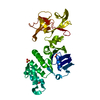
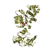
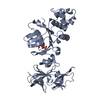
- Assembly
| Deposited unit | 
| ||||||||
|---|---|---|---|---|---|---|---|---|---|
| 1 |
| ||||||||
| Unit cell |
|
-Components
| #1: Protein | Mass: 19823.846 Da / Num. of mol.: 2 / Fragment: DNA binding domain: UNP residues 1-162 Source method: isolated from a genetically manipulated source Source: (gene. exp.) Listeria monocytogenes (bacteria) / Gene: lmo0674, mogR / Production host: #2: DNA chain | | Mass: 4590.046 Da / Num. of mol.: 1 / Source method: obtained synthetically / Details: flaA promoter sequence #3: DNA chain | | Mass: 4581.033 Da / Num. of mol.: 1 / Source method: obtained synthetically / Details: flaA promoter sequence #4: Water | ChemComp-HOH / |  Mass: 18.015 Da / Num. of mol.: 220 / Source method: isolated from a natural source / Formula: H2O Mass: 18.015 Da / Num. of mol.: 220 / Source method: isolated from a natural source / Formula: H2O |
|---|
-Experimental details
-Experiment
| Experiment | Method: X-RAY DIFFRACTION / Number of used crystals: 1 |
|---|
- Sample preparation
| Crystal | Density Matthews: 2.58 Å3/Da / Density % sol: 52.41 % | ||||||||||||||||||||
|---|---|---|---|---|---|---|---|---|---|---|---|---|---|---|---|---|---|---|---|---|---|
| Crystal grow | Temperature: 298 K / Method: vapor diffusion, hanging drop / pH: 6 Details: 100 mM 2-(N-morpholino)ethanesulfonic acid (MES) pH 6.0, 10-15% w/v PEG 4000, VAPOR DIFFUSION, HANGING DROP, temperature 298.0K | ||||||||||||||||||||
| Components of the solutions |
|
-Data collection
| Diffraction | Mean temperature: 100 K |
|---|---|
| Diffraction source | Source: SYNCHROTRON / Site: APS  / Beamline: 24-ID-C / Wavelength: 0.979 Å / Beamline: 24-ID-C / Wavelength: 0.979 Å |
| Detector | Type: ADSC QUANTUM 315 / Detector: CCD / Date: Mar 22, 2007 |
| Radiation | Monochromator: Si(111) / Protocol: SINGLE WAVELENGTH / Monochromatic (M) / Laue (L): M / Scattering type: x-ray |
| Radiation wavelength | Wavelength: 0.979 Å / Relative weight: 1 |
| Reflection | Resolution: 1.74→50 Å / Num. all: 49987 / Num. obs: 48556 / % possible obs: 97.1 % / Observed criterion σ(I): 2 |
| Reflection shell | Resolution: 1.74→1.8 Å / % possible all: 64.8 |
- Processing
| Software |
| ||||||||||||||||||
|---|---|---|---|---|---|---|---|---|---|---|---|---|---|---|---|---|---|---|---|
| Refinement | Method to determine structure: SAD / Resolution: 1.75→50 Å / σ(F): 0 / Stereochemistry target values: Engh & Huber
| ||||||||||||||||||
| Solvent computation | Bsol: 39.534 Å2 / ksol: 0.35 e/Å3 | ||||||||||||||||||
| Displacement parameters |
| ||||||||||||||||||
| Refinement step | Cycle: LAST / Resolution: 1.75→50 Å
| ||||||||||||||||||
| Refine LS restraints |
| ||||||||||||||||||
| LS refinement shell | Resolution: 1.75→1.83 Å / Total num. of bins used: 10
|
